At a Glance
| Factor | Detail |
|---|---|
| What it is | Selective autophagy targeting dysfunctional mitochondria |
| Why it matters | Prevents ROS accumulation, preserves ATP output, protects neurons |
| Declines with age | Measurably impaired by age 40; sharply worse after 60 |
| Top activators | Fasting ≥16 h, aerobic exercise, urolithin A, spermidine, NAD+ precursors |
| Top blockers | Chronic excess calories, sedentary lifestyle, mTOR over-activation, alcohol |
| Clinical relevance | Alzheimer’s, Parkinson’s, metabolic syndrome, sarcopenia all share impaired mitophagy |
Mitochondria are the only organelles with their own DNA — a relic of their ancient bacterial origin. That independent genome makes them powerful, but also vulnerable: mitochondrial DNA (mtDNA) lacks the repair machinery of nuclear DNA and sits just nanometres from the reactive oxygen species (ROS) that mitochondria themselves generate. Over a lifetime, damage accumulates. Mitochondria become less efficient, leak more ROS, and — critically — begin to trigger the same inflammatory and apoptotic cascades they were supposed to prevent.
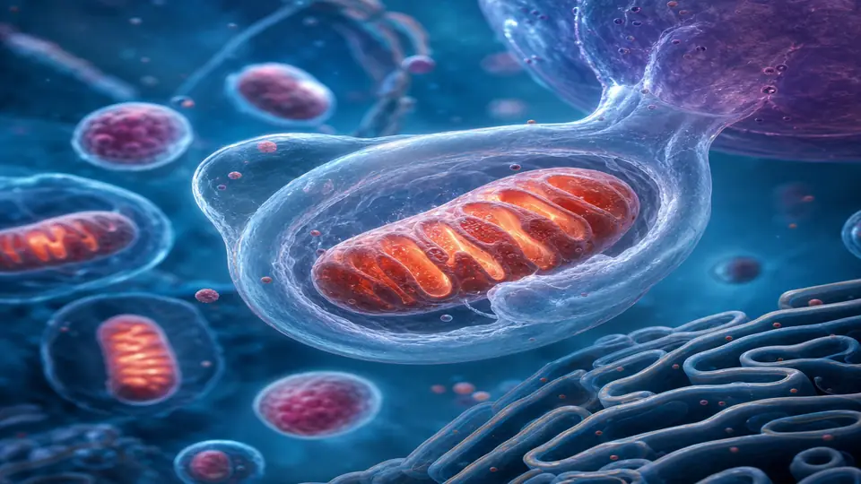
The cellular answer to this problem is mitophagy: a selective form of autophagy that identifies, engulfs, and recycles damaged mitochondria before they can poison the cell. When mitophagy works well, the mitochondrial network stays young and high-performing. When it fails — as it reliably does with age, metabolic excess, and sedentary living — the cell accumulates a population of broken, ROS-spewing organelles that accelerate every hallmark of aging.
This article covers what mitophagy is, why it declines, the clinical consequences when it does, and the interventions with the strongest evidence for restoring it.
What Mitophagy Is (and How It Differs from General Autophagy)
General autophagy is the cell’s bulk recycling programme — a non-selective process that degrades cytoplasmic contents during nutrient scarcity. Mitophagy is its more precise cousin. It uses a dedicated signalling cascade to tag only dysfunctional mitochondria for removal, leaving healthy ones intact.
The canonical pathway runs through two proteins: PINK1 (PTEN-induced kinase 1) and Parkin, an E3 ubiquitin ligase. In a healthy mitochondrion, PINK1 is continuously imported into the inner membrane and degraded. When the membrane potential (ΔΨm) drops — the hallmark of mitochondrial dysfunction — import halts. PINK1 accumulates on the outer membrane, recruits and activates Parkin, which then ubiquitinates outer-membrane proteins. These ubiquitin tags are recognised by autophagy receptors (NDP52, OPTN, p62), which deliver the mitochondrion to the growing phagophore, forming an autophagosome that fuses with the lysosome for degradation.
A parallel, PINK1/Parkin-independent pathway uses mitochondrial membrane proteins BNIP3 and NIX (and their newer cousin FUNDC1) as direct autophagy receptors — critical during hypoxia and in red blood cell maturation.
Both routes converge on the same outcome: the targeted elimination of mitochondria that are no longer fit for purpose.
Why Mitophagy Declines with Age
Several intersecting mechanisms drive age-related mitophagy failure:
1. Reduced PINK1/Parkin Activity
PINK1 expression falls with age in human brain and muscle tissue. Parkin function is also impaired by S-nitrosylation, a post-translational modification that increases with oxidative stress — the very condition damaged mitochondria produce. The result is a feedback loop: more damaged mitochondria → more ROS → more Parkin inhibition → even less mitophagy.
2. Declining NAD+ Levels
The NAD+-dependent deacetylase SIRT1 and the mitochondria-specific SIRT3 are essential co-regulators of mitophagy. They deacetylate and activate key autophagy proteins including Beclin-1 and LC3. NAD+ levels fall roughly 50% between age 20 and 60 in human tissue, directly impairing this regulatory axis. Studies in aged mice show that NAD+ repletion with NMN or NR restores mitophagy flux and extends healthspan.
3. mTOR Hyper-activation
The nutrient-sensing kinase mTOR is the master inhibitor of autophagy. Chronic caloric excess, insulin resistance, and high branched-chain amino acid intake all hyperactivate mTOR, which phosphorylates and inactivates the ULK1 kinase complex — the initiator of autophagosome formation. In ageing adipose and muscle tissue, mTOR signalling is chronically elevated even in the fasted state, creating a permissive environment for mitochondrial accumulation.
4. Lysosomal Dysfunction
Mitophagy is only as good as the lysosome that degrades the cargo. Lysosomal acidification declines with age (cathepsin activity falls), and the accumulation of undegraded lipofuscin occupies lysosomal volume. This “lysosomal overload” bottlenecks the entire autophagy-mitophagy pipeline regardless of how well upstream signalling functions.
Clinical Consequences of Impaired Mitophagy
The connection between failed mitophagy and disease is no longer theoretical.
Neurodegeneration: Loss-of-function mutations in PINK1 and Parkin cause early-onset Parkinson’s disease — the genetic proof-of-concept that mitophagy failure kills dopaminergic neurons. In sporadic Alzheimer’s, accumulation of damaged mitochondria and defective mitophagy are documented in post-mortem hippocampal tissue, and correlate with tau pathology and cognitive decline severity.
Metabolic syndrome: In skeletal muscle, accumulated dysfunctional mitochondria reduce oxidative capacity and produce excess ceramides and diacylglycerol, both of which cause insulin resistance independent of caloric intake. Restoring mitophagy in diet-induced obese mice improves insulin sensitivity without caloric restriction.
Cardiovascular disease: Cardiac mitophagy is required to clear mitochondria damaged during ischaemia-reperfusion. In patients with heart failure, PINK1 expression is reduced, Parkin translocation is impaired, and damaged mitochondria accumulate in cardiomyocytes — contributing to the energetic deficits and contractile dysfunction characteristic of the failing heart.
Sarcopenia: Type II muscle fibre loss with age directly correlates with impaired mitophagy, mitochondrial fragmentation, and reduced oxidative phosphorylation capacity. Muscle-specific Parkin knockout in mice accelerates age-related strength loss.
Chronic fatigue: In patients with myalgic encephalomyelitis/chronic fatigue syndrome (ME/CFS), PBMCs show elevated mitochondrial ROS, reduced ΔΨm, and impaired mitophagy flux — consistent with a toxic accumulation of dysfunctional mitochondria as a driver of the characteristic post-exertional malaise.
Evidence-Based Strategies to Activate Mitophagy
Intermittent and Prolonged Fasting
Fasting is the most potent physiological mitophagy activator. Within 12–16 hours of caloric restriction, falling insulin and glucose levels suppress mTOR and activate AMPK, which phosphorylates and activates ULK1. Mitophagy flux is measurable in human PBMCs after 24-hour fasting, and prolonged fasting (72 h) produces a robust “mitochondrial reset” in muscle and liver.
Practically: a 16:8 intermittent fasting window maintains chronic mitophagy above baseline. A monthly 3–5 day fasting-mimicking diet (800–1,100 kcal from complex carbohydrate and healthy fat) produces a deeper reset with clinical evidence of improved mitochondrial biogenesis markers on refeeding.
Aerobic Exercise and Zone 2 Training
Exercise-induced mitophagy operates via a distinct pathway: calcium release during contraction activates CAMKK2, which activates AMPK, and exercise-induced ROS directly stabilise PINK1 on depolarised mitochondria. Critically, exercise also upregulates PGC-1α, the master regulator of mitochondrial biogenesis — creating a net turnover cycle where damaged mitochondria are cleared and fresh ones are made.
Zone 2 training (60–70% VO2max, conversational pace, 150+ min/week) is most consistently associated with mitophagy induction in human skeletal muscle biopsies. High-intensity interval training drives greater acute mitophagy but with more recovery demands.
Urolithin A
Urolithin A is the gut microbial metabolite of ellagitannins found in pomegranates, berries, and walnuts. It is the only orally available compound with direct, clinically validated evidence for mitophagy induction in humans. In a 2019 Nature Metabolism RCT (n=60), supplemental urolithin A (500–1,000 mg/day for 4 weeks) increased mitophagy gene expression in muscle biopsies and improved mitochondrial gene signatures in blood. A 2022 follow-up showed improved muscle endurance in older adults.
The challenge: only ~40% of adults harbour the gut bacteria (mainly Gordonibacter species) capable of converting ellagitannins to urolithin A. Supplemental urolithin A bypasses this conversion bottleneck. Clinical dose: 500–1,000 mg/day with food.
Spermidine
The polyamine spermidine activates autophagy and mitophagy by inhibiting EP300, a histone acetyltransferase that epigenetically suppresses autophagy genes. In aged mice, oral spermidine supplementation extends lifespan, restores mitophagy flux in cardiac tissue, and improves diastolic function. Human observational data show higher dietary spermidine intake (wheat germ, aged cheese, mushrooms, legumes) correlating with reduced all-cause mortality and lower rates of cognitive decline.
Supplemental dose: 1–5 mg/day. Higher dietary sources (wheat germ provides ~0.6 mg/g) are the most accessible form. Food-based spermidine appears bioavailable and safe across all age groups studied.
NAD+ Precursors (NMN and NR)
NAD+ is required for SIRT1-mediated deacetylation of autophagy and mitophagy proteins. In aged animals, NMN (250–500 mg/day equivalent) and NR (250–1,000 mg/day) restore mitophagy gene expression and improve mitochondrial function across multiple tissues. Human data remain less robust for the mitophagy endpoint specifically, though NMN does demonstrably raise blood NAD+ levels and improve muscle insulin sensitivity in a Phase I trial.
The combination of NAD+ precursors with a mitophagy activator (e.g., urolithin A + NMN) may have additive benefit by simultaneously removing dysfunctional mitochondria and restoring the NAD+ signalling needed to regulate the process.
Rapamycin (Low-Dose, Intermittent)
Rapamycin’s lifespan-extending effect in mammals is largely attributed to mTOR inhibition and consequent autophagy/mitophagy activation. Weekly low-dose rapamycin (0.5–1 mg/week) is increasingly explored in healthy human longevity protocols, with favourable safety profiles in short-term studies. Given its immunosuppressive potential at higher doses, clinical supervision is essential. This is not a first-line intervention, but it has the strongest mechanistic case for chronic mTOR inhibition combined with a fasting window.
What to Avoid: Common Mitophagy Blockers
| Block | Mechanism |
|---|---|
| Chronic caloric excess | Sustained mTOR activation |
| Alcohol (>2 drinks/day) | Impairs PINK1 stability, lysosomal acidification |
| Sedentary lifestyle | Reduces AMPK activation, PGC-1α |
| High-dose antioxidants pre-workout | Scavenge the ROS needed to stabilise PINK1 on damaged mitochondria |
| Late-night eating | Compresses the overnight fasting window below the mitophagy threshold |
| Chronic stress / cortisol excess | mTOR activation via PI3K-Akt; also suppresses TFEB (lysosome biogenesis) |
A notable clinical finding: high-dose vitamin C and vitamin E supplementation before exercise blunts exercise-induced mitophagy signalling, consistent with animal data. The timing of antioxidants matters — taking them several hours after training preserves the signalling benefit while still managing oxidative burden.
A Practical Mitophagy Protocol
For patients without contraindications, the following approach covers all major activation pathways:
- Daily fasting window ≥16 hours (finish dinner by 7 pm, break fast at 11 am)
- Zone 2 aerobic exercise 3–4 × per week, 45–60 min per session
- Urolithin A 500 mg/day with the first meal
- Spermidine 1–5 mg/day from wheat germ or supplemental form
- NMN 250–500 mg/day in the morning
- Avoid antioxidant supplements within 2 hours of training
- Monthly 3-day fasting-mimicking diet for a deeper mitochondrial reset
This is not a stack for everyone. Patients with advanced sarcopenia, eating disorders, or active immune conditions require modified protocols. Always assess muscle mass, inflammatory markers, and metabolic baseline before prescribing fasting durations beyond 16 hours.
Related Articles
- Autophagy: The Science of Cellular Recycling — the broader autophagy framework that mitophagy operates within
- NAD+ Supplement Guide: NMN, NR, and IV NAD — detailed breakdown of NAD+ precursor options and dosing
- Urolithin A: The Gut-Derived Longevity Molecule — deep dive into the clinical evidence for urolithin A
- Zone 2 Training and Longevity — how aerobic base training drives mitochondrial health
- Mitochondria and Cellular Energy — foundational overview of mitochondrial biology and its role in ageing
References
- Pickrell AM, Youle RJ. The roles of PINK1, parkin, and mitochondrial fidelity in Parkinson’s disease. Neuron. 2015;85(2):257–273. PMID 25611507
- Ryu D, et al. Urolithin A induces mitophagy and prolongs lifespan in C. elegans and increases muscle function in rodents. Nature Medicine. 2016;22(8):879–888. PMID 27400265
- Andreux PA, et al. The mitophagy activator urolithin A is safe and induces a molecular signature of improved mitochondrial and cellular health in humans. Nature Metabolism. 2019;1(6):595–603. PMID 32694810
- Eisenberg T, et al. Cardioprotection and lifespan extension by the natural polyamine spermidine. Nature Medicine. 2016;22(12):1428–1438. PMID 27841876
- Mouchiroud L, et al. The NAD+/sirtuin pathway modulates longevity through activation of mitochondrial UPR and FOXO signaling. Cell. 2013;154(2):430–441. PMID 23870130
- Laker RC, et al. Ampk phosphorylation of Ulk1 is required for targeting of mitochondria to lysosomes in exercise-induced mitophagy. Nature Communications. 2017;8(1):548. PMID 28916822
- Mills KF, et al. Long-term administration of nicotinamide mononucleotide mitigates age-associated physiological decline in mice. Cell Metabolism. 2016;24(6):795–806. PMID 28068222