The patient sitting across from me had seen eleven specialists. Depression, cognitive decline, and chronic bloating had consumed the better part of three years. Every organ had been evaluated in isolation. No one had connected the dots between the gut and the brain.
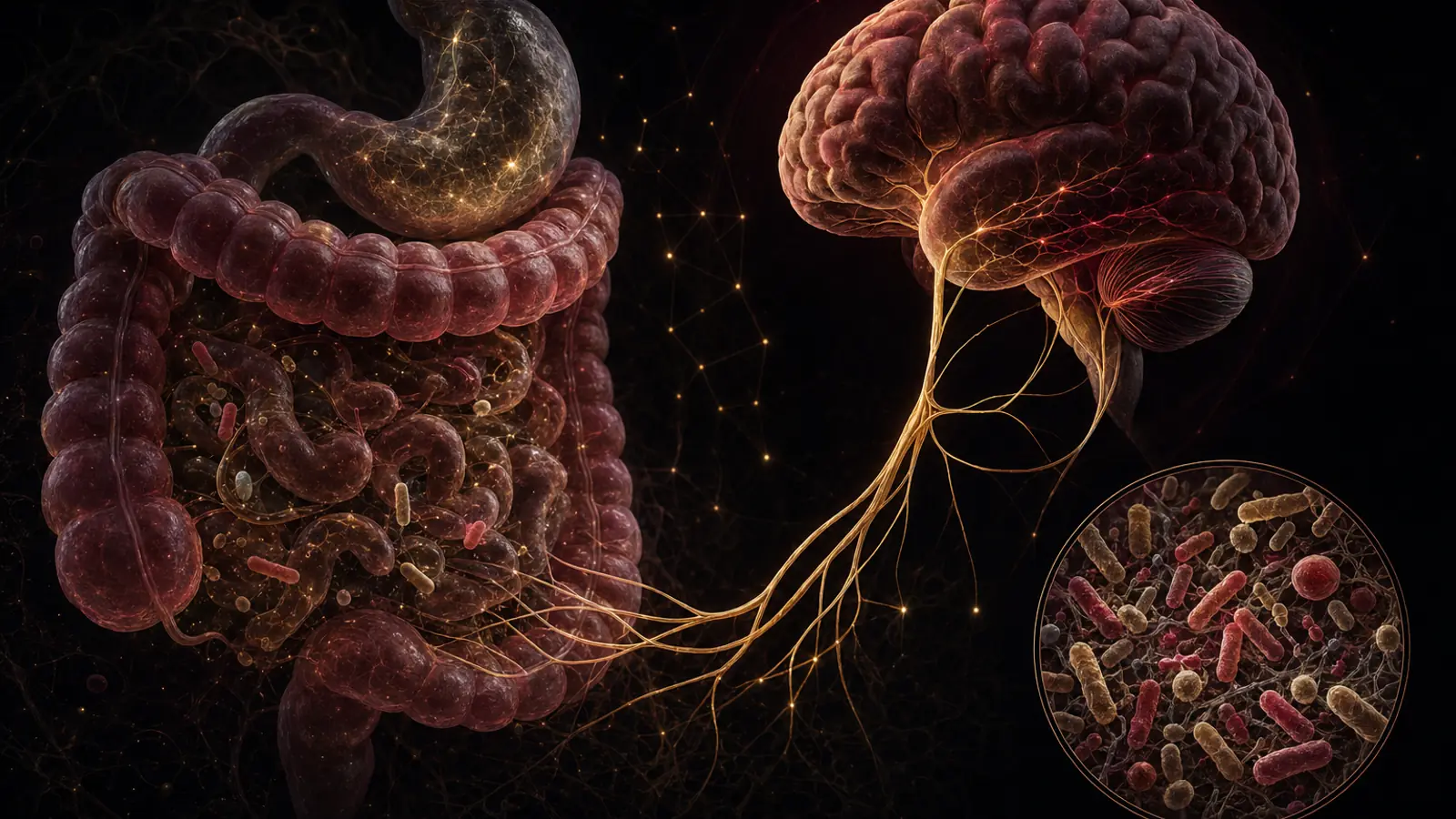
That scenario is more common than it should be. The gut-brain axis is the two-way communication network linking the enteric nervous system (the gut’s own nervous system), the immune system and the central nervous system. It is one of the most clinically consequential discoveries of the past two decades, and it changes how we approach anxiety, depression, brain fog and autoimmune conditions as well as digestive disorders.
At a Glance
| Feature | Key Point |
|---|---|
| What it is | Bidirectional signalling network between gut microbiome, enteric nervous system, immune system, and brain |
| Primary highway | Vagus nerve (carries ~80% of signals gut-to-brain) |
| Key microbial products | Short-chain fatty acids, neurotransmitter precursors, lipopolysaccharide (LPS) |
| Disruption patterns | Dysbiosis, leaky gut, neuroinflammation, mood disorders |
| Restoration targets | Microbiome diversity, mucosal barrier integrity, vagal tone, systemic inflammation |
| Clinical timeline | Measurable microbiome shifts within 4–8 weeks; symptom improvement typically 8–16 weeks |
What Is the Gut-Brain Axis?
The gut-brain axis is a network rather than a single anatomical structure, with four principal components:
1. The enteric nervous system (ENS). Often called the “second brain,” the ENS contains roughly 500 million neurons embedded in the lining of the gastrointestinal tract. It operates semi-autonomously, regulating peristalsis, secretion, and local immune responses without waiting for instructions from the brain.
2. The vagus nerve. This cranial nerve is the primary conduit between the gut and the central nervous system. Approximately 80% of vagal fibers are afferent, meaning they carry signals upward from the gut to the brainstem. The brain receives a constant stream of information from the intestinal environment.
3. The hypothalamic-pituitary-adrenal (HPA) axis. Stress hormones alter gut motility, permeability, and microbial composition. The gut, in turn, modulates cortisol signalling. This creates a feedback loop that can amplify or dampen the stress response depending on the state of the microbiome.
4. The gut microbiome itself. The roughly 38 trillion microorganisms residing in the human gut produce neurotransmitters, metabolites, and inflammatory signals that reach the brain through multiple routes: directly via the vagus nerve, indirectly via the bloodstream, and through immune cell trafficking across the gut-blood-brain barrier interface.
The Microbial Machinery: How Bacteria Talk to Your Brain
The gut microbiome exerts influence over the central nervous system through several well-characterised mechanisms.
Neurotransmitter Production
Approximately 90–95% of the body’s total serotonin is produced in the gut, not the brain. Enterochromaffin cells synthesize serotonin in response to microbial signals, particularly from Lactobacillus and Bifidobacterium species. This gut-derived serotonin cannot cross the blood-brain barrier, but it plays a major role in peristalsis and nausea signalling and, via vagal afferents, influences central mood regulation indirectly.
GABA, the brain’s primary inhibitory neurotransmitter, is also produced by gut bacteria. Specific strains of Lactobacillus rhamnosus have been shown in animal models to alter GABA receptor expression in the brain, with downstream effects on anxiety behaviour.
Short-Chain Fatty Acids
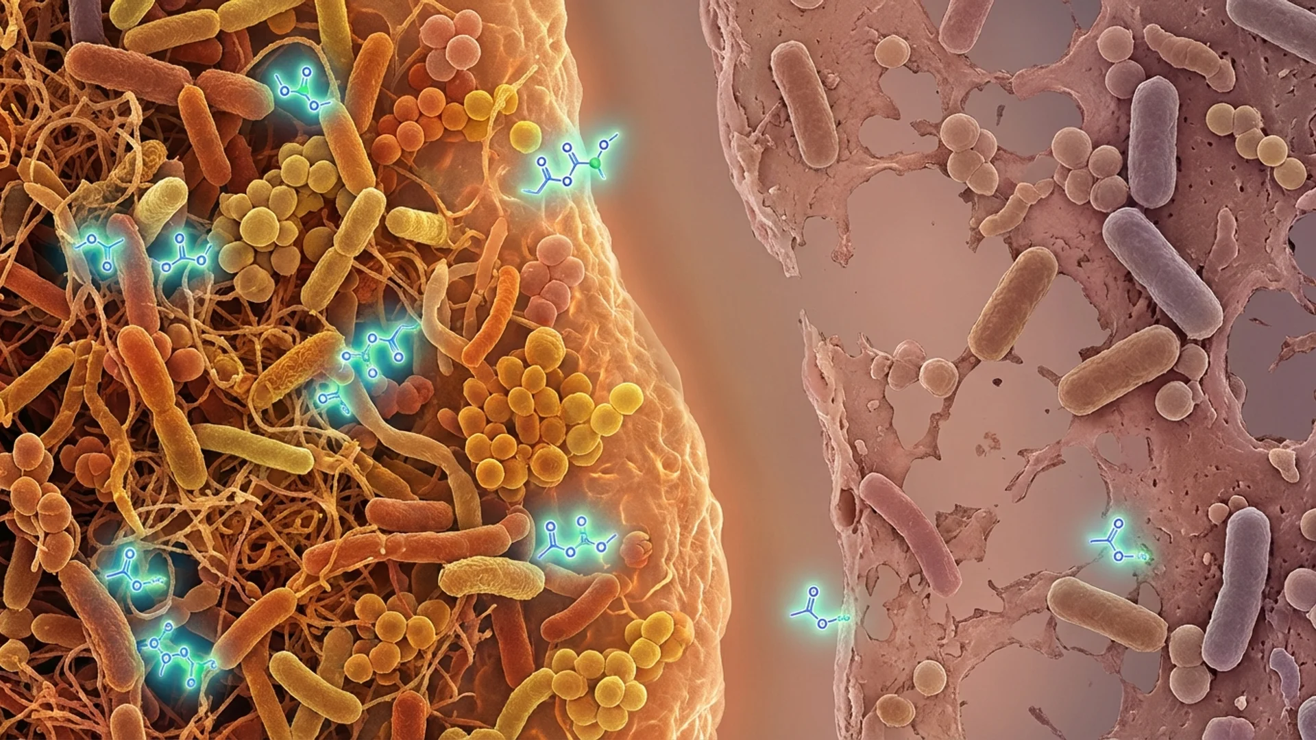
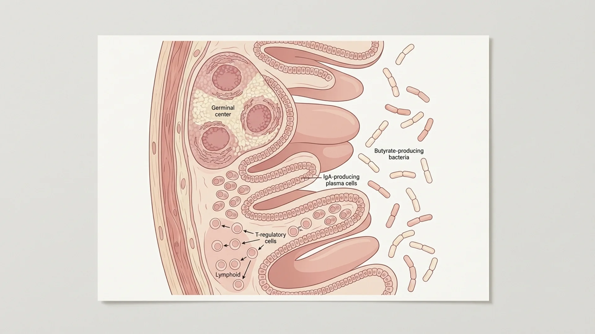
When commensal bacteria ferment dietary fibre, they produce short-chain fatty acids (SCFAs), primarily butyrate, propionate and acetate. Butyrate is the primary fuel source for colonocytes and plays a central role in maintaining the intestinal mucosal barrier. Beyond the gut, SCFAs cross the blood-brain barrier and exert direct anti-inflammatory effects on microglia, the brain’s resident immune cells. Low butyrate production, a consistent finding in dysbiotic microbiomes, correlates clinically with neuroinflammation, depression and cognitive decline.
Lipopolysaccharide and Intestinal Permeability
Gram-negative bacteria shed lipopolysaccharide (LPS), a potent immune activator, as a byproduct of their cell wall turnover. Under normal conditions, the mucosal barrier prevents LPS from entering systemic circulation. When intestinal permeability increases (“leaky gut”), LPS translocates into the bloodstream and triggers a chronic, low-grade inflammatory state. Elevated serum LPS levels have been documented in major depressive disorder, Alzheimer’s disease and Parkinson’s disease, reproducibly across multiple independent cohorts.
The mucus-layer bacterium Akkermansia muciniphila acts as a primary gatekeeper against this translocation: when present at healthy abundance it reinforces tight junction proteins and keeps LPS confined to the lumen. A 2019 Nature Medicine RCT confirmed that pasteurized Akkermansia supplementation reduced both insulin resistance and systemic inflammation markers in overweight volunteers, an effect likely mediated by this barrier-sealing mechanism. For the full clinical picture, see Akkermansia muciniphila: evidence-based guide to the gut barrier strain.
When the Axis Breaks Down: Clinical Patterns
Gut-brain axis dysfunction rarely announces itself with a single dramatic symptom. More often, it presents as a cluster of seemingly unrelated complaints that practitioners evaluate separately and treat, ineffectively, in isolation.
The Brain Fog–Bloating–Fatigue Triad
This is the most common presentation I encounter in practice. Patients report cognitive slowing, post-meal fatigue, and variable bowel symptoms. On microbiome testing, the typical finding is low microbial diversity, overgrowth of gram-negative proteobacteria, and deficient SCFA-producing species. Inflammatory markers, particularly hsCRP (high-sensitivity C-reactive protein) and IL-6, are often mildly but persistently elevated.
The mechanism is straightforward: LPS-driven systemic inflammation activates microglial cells in the prefrontal cortex, increasing inflammatory cytokine production. The brain fog has a measurable biochemical basis, even when it is dismissed as psychological. We discuss the full interpretation of these inflammatory markers in our functional medicine lab interpretation guide.
Mood Disorders and Dysbiosis
The association between gut dysbiosis and depression is bidirectional. Stress reduces microbial diversity by elevating cortisol and altering gut motility. Reduced diversity impairs SCFA production and increases intestinal permeability, which drives inflammation, which worsens mood and closes the loop.
Clinically, I have observed that patients with treatment-resistant depression who have not responded to conventional antidepressants frequently have significant dysbiosis on GI-MAP or organic acid testing. Addressing the gut does not replace psychiatric care, but it often removes a significant driver of the inflammatory burden that makes conventional treatment less effective.
Neurological Conditions and Microbiome Signatures
The data linking microbiome composition to neurodegenerative disease is compelling. In Parkinson’s disease, gut symptoms such as constipation and altered motility precede motor symptoms by up to a decade in many patients. The pathological protein alpha-synuclein has been found in enteric neurons before its detection in the brain, suggesting a gut-to-brain propagation pathway via the vagus nerve.
In the patients I see with chronic Lyme disease, post-COVID neurological symptoms, and mold-related illness, gut dysbiosis is nearly universal. The intestinal barrier disruption and LPS translocation these conditions generate amplifies the central neuroinflammation that drives cognitive symptoms. In these cases, restoring gut integrity is foundational care. This intersection of gut health and Lyme-related neurological symptoms is explored further in our article on Lyme disease and gut dysbiosis.
Assessing the Gut-Brain Axis in Clinical Practice
A thorough evaluation requires looking beyond standard gastroenterology panels.
GI-MAP (Quantitative PCR stool analysis) provides DNA-based quantification of bacteria, parasites, viruses, and fungi, along with markers of intestinal inflammation (calprotectin, secretory IgA) and mucosal integrity (zonulin, beta-glucuronidase). This is my first-line investigation for any patient with concurrent gut and neurological symptoms.
Organic acid testing (OAT) captures microbial metabolite production, including markers of yeast overgrowth (arabinose), SCFA pathways, and mitochondrial function. It shows what the gut microbiome is producing, which complements a list of the organisms present.
Serum LPS-binding protein and zonulin provide direct evidence of intestinal permeability and endotoxin translocation. Zonulin is particularly useful as a repeatable, non-invasive marker of barrier integrity over time.
HRV (heart rate variability) serves as a functional proxy for vagal tone. Low HRV correlates with reduced vagal modulation of the gut and brain, and it is a modifiable biomarker with direct therapeutic implications.
Restoring the Gut-Brain Axis: Evidence-Based Protocols
Restoration requires a phased approach. Sequence matters.
Phase 1: Remove and Reduce (Weeks 1–4)
Before rebuilding, identify and address active drivers of dysbiosis. Pathogens, small intestinal bacterial overgrowth (SIBO), and yeast overgrowth need to be addressed before attempting microbiome restoration. Attempting to reseed a dysbiotic environment rarely works. Targeted antimicrobial botanicals such as berberine, oregano oil and allicin are commonly used in this phase based on GI-MAP findings, with concurrent attention to diet quality and removal of dysbiosis-promoting ultra-processed foods.
Phase 2: Restore Barrier Integrity (Weeks 2–8, overlapping)
Mucosal barrier restoration begins with butyrate-producing nutrients. Clinically validated approaches include:
- Sodium butyrate (300–600 mg twice daily with meals): Direct provision of the primary colonocyte fuel and tight-junction regulator
- L-glutamine (5–15 g daily): Substrate for enterocyte turnover and tight-junction protein synthesis: for dosing protocols by clinical context, see L-Glutamine for Gut Health
- Zinc carnosine (75 mg twice daily): Well-studied in mucosal repair; superior bioavailability for gut-wall support compared to standard zinc forms
- Bovine colostrum (10–20 g/day): Provides IgG, lactoferrin, and TGF-β to directly reinforce tight junctions and support mucosal IgA secretion: see bovine colostrum clinical review
- Colostrum or immunoglobulin concentrates: Provide secretory IgA and growth factors that support mucosal immunity
Dietary fibre is essential, and the type matters. Resistant starch (green banana, cooked and cooled potato, inulin) specifically feeds butyrate-producing Firmicutes species such as Faecalibacterium prausnitzii and Roseburia intestinalis, which are the primary producers of mucosal-protective SCFAs.
Phase 3: Reseed and Diversify (Weeks 4–16)
Targeted probiotic supplementation is most effective once the dysbiotic load has been reduced and the mucosal environment is less hostile. Multi-strain formulations including Lactobacillus plantarum, L. rhamnosus, Bifidobacterium longum, and B. infantis have the strongest evidence base for gut-brain axis modulation. Turkey tail mushroom (Trametes versicolor) is a useful adjunct here: its polysaccharopeptide (PSP) fraction has demonstrated significant bifidogenic effects in randomised trials, selectively increasing Bifidobacterium and Lactobacillus populations. Turkey tail’s oncology and microbiome data are covered in our overview of medicinal mushrooms and their immune mechanisms. Fermented foods such as kefir, kimchi, sauerkraut and miso, when tolerated, accelerate microbial diversification.
Psychobiotics, specific probiotic strains shown to influence mental health endpoints, deserve mention. A 2019 study in Beneficial Microbes demonstrated that a Lactobacillus helveticus/Bifidobacterium longum combination reduced self-reported anxiety and cortisol output in stressed adults. Effects were modest but consistent, and the proposed mechanism, reduced HPA (hypothalamic-pituitary-adrenal) axis reactivity, fits the broader gut-brain axis literature.
Phase 4: Support Vagal Tone
Gut microbiome restoration and vagal tone enhancement are complementary, not sequential. Practical vagal tone interventions with documented physiological effects include:
- Diaphragmatic breathing: 5–6 breath cycles per minute for 10–20 minutes daily; increases high-frequency HRV within 4 weeks
- Cold water facial immersion: Activates the diving reflex and parasympathetic response
- Humming and gargling: Directly stimulates vagal efferents via the pharyngeal branch
- Aerobic exercise at zone 2 intensity: Increases basal vagal tone with consistent training
In our clinic, we frequently combine gut restoration protocols with photobiomodulation and neurofeedback for patients presenting with significant neuroinflammation and vagal dysfunction. The neurological dimension of these approaches is covered in depth in our photobiomodulation guide and the vagus nerve article.
The Integrative Perspective
The gut-brain axis is one of the clearest examples of why organ-siloed medicine falls short for complex chronic conditions. The intestinal environment continuously influences brain chemistry, immune activation and stress reactivity, and those systems in turn shape the gut.
In clinical practice, this means that patients with persistent neurological or psychiatric symptoms deserve a thorough gut assessment, and that patients presenting with chronic gut disorders should be screened for neurological and mood co-morbidities. Both often come from the same disrupted biology, so sending the patient to two separate specialists can miss the connection.
The good news is that the gut is among the most therapeutically responsive systems in the body. Measurable microbiome changes occur within days of dietary intervention; mucosal barrier markers respond within weeks; and clinical symptom improvement typically follows within one to four months of a structured protocol. That is a faster response timeline than most pharmaceutical interventions for depression or cognitive decline.
Our approach begins with diagnostics that show what is dysregulated in the individual patient, before any protocol is chosen. If you recognise the pattern described here, a full gut-brain axis evaluation is a reasonable starting point. More detail on our diagnostic approach can be found in our gut microbiome testing overview and the GI-MAP vs standard culture comparison.
Related Articles
- Gut Microbiome Testing: What the Results Actually Mean
- The Vagus Nerve: Inflammation, Immunity, and How to Tone It
- Lyme Disease and Gut Dysbiosis: A Hidden Connection
- GI-MAP vs Standard Stool Culture: Which Test Do You Need?
- Post-COVID Brain Fog: Mechanisms and Treatment Approaches
References
-
Cryan JF, O’Riordan KJ, Cowan CSM, et al. The Microbiota-Gut-Brain Axis. Physiol Rev. 2019;99(4):1877–2013. PMID 31460832
-
Carabotti M, Scirocco A, Maselli MA, Severi C. The gut-brain axis: interactions between enteric microbiota, central and enteric nervous systems. Ann Gastroenterol. 2015;28(2):203–209. PMID 25830558
-
Dinan TG, Cryan JF. Gut instincts: microbiota as a key regulator of brain development, ageing and neurodegeneration. J Physiol. 2017;595(2):489–503. PMID 27641441
-
Mayer EA, Knight R, Mazmanian SK, Cryan JF, Tillisch K. Gut microbes and the brain: paradigm shift in neuroscience. J Neurosci. 2014;34(46):15490–15496. PMID 25392516
-
Kelly JR, Kennedy PJ, Cryan JF, Dinan TG, Clarke G, Hyland NP. Breaking down the barriers: the gut microbiome, intestinal permeability and stress-related psychiatric disorders. Front Cell Neurosci. 2015;9:392. PMID 26528128
-
Pellegrini C, Antonioli L, Colucci R, Blandizzi C, Fornai M. Interplay among gut microbiota, intestinal mucosal barrier and enteric neuroimmune system: a common path to neurodegenerative diseases? Acta Neuropathol. 2018;136(3):345–361. PMID 29797112
-
Vuong HE, Yano JM, Fung TC, Hsiao EY. The Microbiome and Host Behavior. Annu Rev Neurosci. 2017;40:21–49. PMID 28301775