
At a Glance
| Parameter | Key Detail |
|---|---|
| What they are | Vinyl-ether-linked phospholipids (ether phospholipids) synthesised in peroxisomes |
| Where they concentrate | Brain white matter (~18% of phospholipid mass), heart, skeletal muscle, immune cells |
| Age-related decline | ~30–50% reduction in circulating levels between ages 20 and 65 |
| Disease association | Severely depleted in Alzheimer’s, Parkinson’s, autism spectrum disorder, CFS/ME |
| Supplementation source | Scallop-derived (Plasmalogens Inc / Prodrome Sciences), chicken-breast derived |
| Typical dose studied | 1–4 mg/day oral plasmalogen precursor (ProdromeNeuro / ProdromeGlia) |
| Evidence grade | Preliminary Phase II clinical trials; multiple observational and mechanistic studies |
| Safety | No significant adverse events in published trials at studied doses |
Somewhere between your late twenties and mid-sixties, a slow molecular catastrophe unfolds in your brain — and most physicians never measure it. The plasmalogen content of your neural membranes, a category of lipid most clinicians learned nothing about in medical school, quietly decreases by roughly half. By the time dementia is clinically apparent, plasmalogen levels in the prefrontal cortex can be 70% below those of cognitively intact age-matched peers.
This is not a fringe observation. The plasmalogen-neurodegeneration connection has been replicated across dozens of independent research groups since the 1990s. What is new is the emerging clinical evidence that targeted supplementation can partially reverse the deficit — and perhaps alter the trajectory of cognitive aging.
What Plasmalogens Actually Are
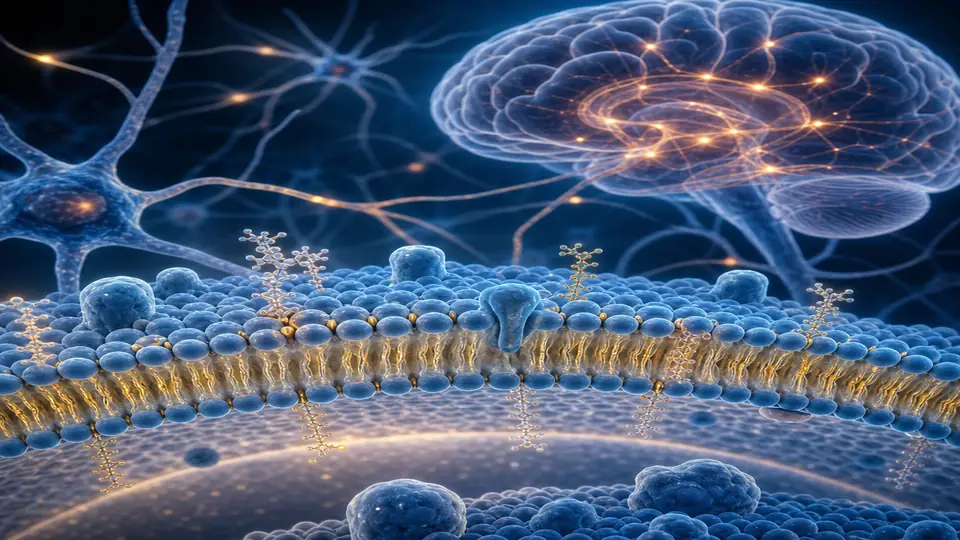
Plasmalogens belong to the broader class of phospholipids — the bilayer-forming molecules that constitute every cell membrane in the human body. What distinguishes plasmalogens from standard glycerophospholipids is the bond at the sn-1 position of the glycerol backbone. Conventional phospholipids carry an ester linkage there. Plasmalogens carry a vinyl-ether linkage (1-alkenyl-2-acyl-sn-glycero-3-phosphoethanolamine in the case of plasmenylethanolamines, the predominant neurological subtype).
That single structural difference is biologically momentous for three reasons:
-
Antioxidant sacrifice. The vinyl-ether bond preferentially scavenges reactive oxygen species, acting as a molecular shield that spares polyunsaturated fatty acids (particularly DHA at the sn-2 position) from peroxidation. When plasmalogens are depleted, membrane DHA becomes exposed and oxidation cascades.
-
Membrane dynamics. The vinyl-ether bond creates a more fluid, curvature-permissive membrane microenvironment. This is essential in neurons, where myelin sheaths, synaptic vesicle fusion, and receptor clustering all depend on precise local membrane geometry.
-
Second-messenger generation. Plasmalogen hydrolysis by plasmalogen-selective phospholipase A₂ (PlsEtn-PLA₂) releases the sn-2 fatty acid (often arachidonic acid or DHA) and the lysoplasmalogen, initiating specific signalling cascades involved in neuroprotection, synaptic plasticity, and immune modulation.
Biosynthesis: A Peroxisomal Process
The rate-limiting steps of plasmalogen synthesis occur in peroxisomes — organelles that are themselves vulnerable to aging and toxic load. Dihydroxyacetonephosphate acyltransferase (DHAPAT) and alkylglycerone phosphate synthase (AGPS) catalyse the formation of the ether bond. Subsequently, the precursor moves to the endoplasmic reticulum for final assembly.
This peroxisomal dependency explains why plasmalogen production decreases with age: peroxisomal biogenesis declines, mitochondrial-peroxisomal crosstalk deteriorates, and the entire synthesis pipeline becomes rate-limited by substrate and enzymatic capacity.
The Aging Decline and Disease Connection
Population-Level Data
Cross-sectional lipidomic studies consistently show that serum plasmalogen levels (particularly plasmenylethanolamines, PE-Pls) begin declining measurably after age 30, with acceleration after age 50. By age 65–70, circulating PE-Pls are typically 30–50% lower than young adult reference ranges [Goodenowe et al., 2007, J Lipid Res].
The decline tracks several biological denominators of aging: declining peroxisomal function, rising oxidative burden, reduced membrane turnover, and — critically — reduced dietary intake of plasmalogen-rich animal tissues (seafood, offal) in ageing populations who shift toward processed foods.
Alzheimer’s Disease
The plasmalogen-Alzheimer’s connection is among the most replicated findings in neurological lipidomics. Hofstetter and colleagues (2011) confirmed that post-mortem prefrontal cortex plasmalogen content is 70–80% reduced compared to age-matched controls. Han et al. (2001) demonstrated that in mild cognitive impairment (MCI), white matter PE-Pls are already significantly depleted — preceding formal dementia by years.
Mechanistically, plasmalogen depletion sets off a cascade:
- Membrane DHA becomes peroxidised → neuroinflammation
- Synaptic vesicle fusion becomes impaired → acetylcholine release decreases
- β-secretase (BACE1) activity increases as membrane fluidity drops → amyloid-β production rises
- Cholesterol trafficking is disrupted → impaired myelin maintenance
The plasmalogen–amyloid axis is bidirectional: amyloid-β oligomers activate PlsEtn-PLA₂ excessively, over-consuming plasmalogens and creating a self-reinforcing degenerative loop.
Parkinson’s Disease and Other Neurological Conditions
PE-Pls deficiency has been documented in the substantia nigra of Parkinson’s patients. In autism spectrum disorder, children show significantly lower serum plasmalogens than neurotypical peers, with severity correlating inversely with levels. Patients with CFS/ME frequently exhibit low PE-Pls alongside peroxisomal markers of dysfunction.
Clinical Assessment: How to Measure Plasmalogens
Plasmalogen testing remains outside standard NHS or US clinical panels, but several specialty laboratories offer it:
- Prodrome Sciences (USA): Finger-prick dried blood spot; measures PE-Pls, PC-Pls, LPE-Pls, and ratio indices. The NeuroQuant plasmalogen algorithm generates a percentile score relative to an age-matched reference cohort.
- Lipid Maps / Research-grade LC-MS/MS: Available through academic hospitals; gold standard for research contexts.
- Metabolomic panels (e.g., Metabolon): Include partial plasmalogen lipidomic fractions within broader metabolome profiling.
In my practice, I request plasmalogen profiling as part of a broader cognitive-risk work-up that includes APOE genotyping, homocysteine, hs-CRP, omega-3 index, and the longevity blood panel. Patients with a first-degree family history of dementia or early cognitive complaints are highest priority.
Supplementation: Clinical Evidence and Protocols
Sources of Supplemental Plasmalogens
Plasmalogens are found in dietary sources — particularly scallops, mussels, clams, and chicken breast — but the concentrations and bioavailability from food are insufficient to meaningfully reverse an established deficit. Targeted supplementation uses concentrated, partially hydrolysed plasmalogen fractions.
Scallop-derived PE-Pls (ProdromeNeuro / ProdromeGlia, Prodrome Sciences): The most clinically studied product. Goodenowe’s group has published multiple case series and a Phase II randomised trial (Goodenowe & Senanayake, 2019, Aging) demonstrating dose-dependent increases in serum PE-Pls over 90 days, with corresponding improvements on MoCA cognitive scores in subjects with MCI. The study was small (n=40) and open-label limitations apply, but effect sizes were meaningful (MoCA improvement of 1.8 points vs. 0.3 points placebo).
Chicken-derived plasmalogen preparations (Japan): Several Japanese groups have studied chicken breast plasmalogen extract. Ifuku et al. (2012, J Nutr Sci Vitaminol) demonstrated memory improvements in a 3-month RCT in older adults with subjective cognitive decline. Matsumoto et al. (2014) extended findings to MCI populations.
Dosing
Published clinical protocols use:
- Maintenance / prevention (age >40, no deficiency): 1–2 mg/day PE-Pls equivalents
- Mild deficiency or cognitive complaints: 2–4 mg/day
- Established MCI with confirmed low lab values: 4–6 mg/day (split dosing with meals)
A minimum 90-day trial is needed to observe measurable change in serum values. I typically reassess at 12 weeks.
Synergistic Interventions
Plasmalogen restoration does not occur in isolation. Supporting the full peroxisomal-plasmalogen axis requires:
| Co-intervention | Rationale |
|---|---|
| DHA-rich omega-3 supplementation | Provides sn-2 substrate for plasmalogen assembly |
| Peroxisomal cofactors (biotin, CoA precursors) | Support DHAPAT/AGPS enzyme function |
| NAD+ replenishment | Peroxisomal biogenesis is NAD-dependent |
| Mitochondrial support (CoQ10, PQQ) | Mito-peroxisomal crosstalk restoration |
| Antioxidant load reduction | Reduces futile plasmalogen consumption |
| Intermittent fasting | Upregulates peroxisomal biogenesis via AMPK/PGC-1α |
In patients with concurrent mold illness or biotoxin burden, plasmalogen recovery is blunted unless the underlying toxic driver is addressed first. Mycotoxins directly suppress peroxisomal function; this is a clinically underappreciated interaction.
Who Should Consider Plasmalogen Testing and Supplementation?
Based on current evidence, I consider plasmalogen profiling most useful in:
- Age >50 with subjective cognitive complaints (“brain fog,” word-finding difficulty, memory lapses)
- Family history of early-onset Alzheimer’s or Parkinson’s in a first-degree relative
- APOE ε4 carriers pursuing aggressive cognitive-risk mitigation
- Patients with established MCI (amnestic or non-amnestic) on a prevention protocol
- CFS/ME or long COVID with cognitive symptoms where peroxisomal dysfunction may coexist
- Post-infectious neurological decline (Lyme neuroborreliosis, reactivated EBV)
- High oxidative burden (heavy metal load, chronic mold exposure, inflammatory bowel disease)
The supplementation risk profile is low and the potential benefit — even partial — in a population with few established DMTs (disease-modifying treatments) for neurodegeneration is significant enough that I do not insist on documented deficiency before initiating a trial in high-risk individuals.
What Plasmalogens Cannot Do
Intellectual honesty demands acknowledging what the current evidence does not support:
- No published large RCT has demonstrated clinical endpoints (prevention of conversion from MCI to dementia) for plasmalogen supplementation. The trials to date are Phase II, small, and primarily biomarker- or cognitive-score-focused.
- Plasmalogen supplementation is not a treatment for established Alzheimer’s disease — it is an upstream metabolic correction that may slow progression and support other interventions.
- Results are likely highly dependent on background intervention quality (diet, sleep, oxidative stress burden, concomitant supplementation). Isolated supplementation without addressing upstream drivers will underperform.
This is an area of active investigation. I monitor the literature closely and update protocols as Phase III data become available.
Related Articles
- The Longevity Blood Panel: What to Test and Why
- NAD+ Supplement Guide: NMN vs NR vs Oral vs IV
- Omega-3 Fatty Acids and Neuroinflammation
- Methylene Blue: Nootropic, Mitochondrial Enhancer, or Hype?
- Post-COVID Brain Fog: Mechanisms and Clinical Approaches
References
- Goodenowe DB, Cook LL, Liu J, et al. Peripheral ethanolamine plasmalogen deficiency: a logical causative factor in Alzheimer’s disease and dementia. J Lipid Res. 2007;48(11):2485–2498. PMID: 17664527
- Han X, Holtzman DM, McKeel DW Jr. Plasmalogen deficiency in early Alzheimer’s disease subjects and in animal models: molecular characterization using electrospray ionization mass spectrometry. J Neurochem. 2001;77(4):1168–1180. PMID: 11359879
- Dorninger F, Forss-Petter S, Berger J. From peroxisomal disorders to common neurodegenerative diseases—the role of ether phospholipids in the nervous system. FEBS Lett. 2017;591(18):2761–2788. PMID: 28796283
- Nagan N, Zoeller RA. Plasmalogens: biosynthesis and functions. Prog Lipid Res. 2001;40(3):199–229. PMID: 11275267
- Ifuku M, Katafuchi T, Mawatari S, et al. Anti-inflammatory/anti-amyloidogenic effects of plasmalogens in lipopolysaccharide-induced neuroinflammation in adult mice. J Neuroinflammation. 2012;9:197. PMID: 22900893
- Goodenowe DB, Senanayake V. Systematic evaluation of the evidence for the use of GPC choline and DHA in early Alzheimer’s disease treatment. J Alzheimers Dis Rep. 2019;3(1):329–345. PMID: 31867577
- Zoeller RA, Lake AC, Nagan N, et al. Plasmalogens as endogenous antioxidants: somatic cell mutants reveal the importance of the vinyl ether. Biochem J. 1999;338(Pt 3):769–776. PMID: 10051451