
At a Glance
| Feature | Detail |
|---|---|
| Pathogen | TBE virus (Flaviviridae); European, Siberian, Far Eastern subtypes |
| Vector | Ixodes ricinus (Europe), I. persulcatus (Asia) |
| Endemic zones | Central/Eastern Europe, Baltic states, Russia, parts of Asia |
| Incubation | 7–14 days (range 2–28 days) |
| Clinical phases | Phase 1: flu-like (4–10 days) → Phase 2: neurological (30–50% of symptomatic cases) |
| Neurological forms | Meningitis (50%), meningoencephalitis (40%), meningoencephalomyelitis (10%) |
| Diagnosis | IgM/IgG serology; LP in neurological phase |
| Vaccine | Highly effective; recommended for residents and travellers in endemic areas |
| No antiviral | Treatment is supportive; recovery can take months to years |
| Long-term sequelae | Post-encephalitic syndrome in 40–60% of neurological cases |
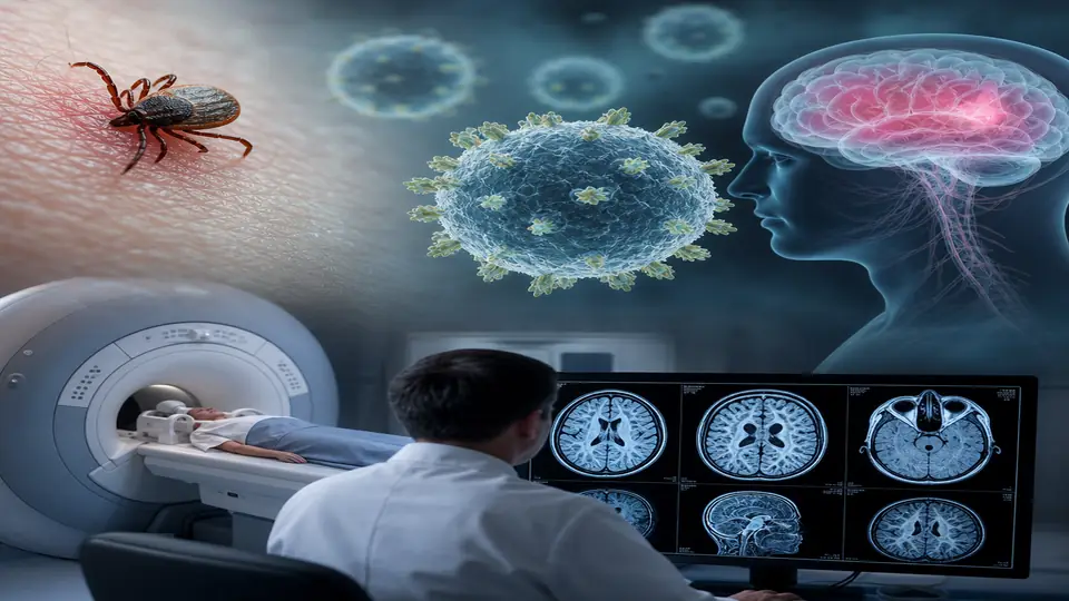
Tick-borne encephalitis is among the most clinically significant viral infections transmitted by ticks in Europe, yet it remains poorly recognised outside endemic medicine. In my clinical practice in Germany — one of the highest-risk countries on the continent — I encounter patients who have received a Lyme disease diagnosis without anyone checking TBE serology, or who are months into post-encephalitic fatigue and cognitive impairment without a coherent recovery plan. This article covers what you need to know: the epidemiology, the diagnostic workup, the biphasic clinical course, and the integrative strategies that can meaningfully support neurological recovery when conventional neurology offers little beyond time.
What Is Tick-Borne Encephalitis?
Tick-borne encephalitis (TBE) is caused by the TBE virus (TBEV), a positive-sense RNA virus in the Flaviviridae family — the same family as dengue, Zika, and West Nile viruses. Three subtypes exist: the European subtype (transmitted by Ixodes ricinus), the Siberian subtype, and the Far Eastern subtype (both transmitted by I. persulcatus). In clinical practice across Germany, Austria, Switzerland, and Scandinavia, the European subtype dominates.
The virus is maintained in a woodland rodent–tick cycle. Humans are incidental dead-end hosts infected through tick bites (the major route) or, less commonly, through unpasteurised dairy products from infected animals. Unlike Lyme disease, TBE cannot be transmitted by a tick that has attached for only a few minutes — TBE virus is present in tick saliva from the moment of attachment, meaning transmission can occur very rapidly.
Epidemiology: A Rising Threat
Reported TBE cases in Europe have increased significantly over the past three decades, driven by climate change expanding tick habitat, increasing outdoor activity, and improved recognition. Germany reports roughly 400–700 cases per year, with Bavaria and Baden-Württemberg as the primary risk regions. Risk areas are expanding northward into previously non-endemic zones including Scandinavia and the Netherlands.
High-risk activities include:
- Walking in forest undergrowth or tall grass below 1,500 m elevation (most risk)
- Camping, hunting, mushroom picking
- Farming and forestry work in endemic areas
- Consuming raw milk (goat, sheep) from endemic farms
Roughly 70% of infections are subclinical or mildly symptomatic. Of those who develop a symptomatic illness, approximately 30–50% progress to the neurological second phase.
The Biphasic Clinical Course
The hallmark of TBE is its biphasic presentation, which distinguishes it from many other encephalitides. Understanding this pattern prevents the common error of reassuring patients that they have recovered when they are actually between phases.
Phase 1: Viraemic Phase (Days 1–8)
After an incubation period of 7–14 days, the viraemic phase presents with:
- Fever (38–40°C), typically abrupt onset
- Severe headache and fatigue
- Myalgia and arthralgia
- Nausea and vomiting
- Occasionally a maculopapular rash
This phase lasts 4–10 days and is clinically indistinguishable from influenza or early Lyme disease. It corresponds to active viraemia and initial immune activation. Most patients (and their physicians) dismiss this as a viral illness and do not seek TBE-specific testing.
Asymptomatic Interval (Days 8–21)
A critical interphase of apparent recovery lasting 1–21 days follows. The patient feels better. This is the viraemia clearing — but in a subset of patients, virus has already seeded the central nervous system.
Phase 2: Neurological Phase (Begins Days 10–21)
The second fever wave heralds CNS involvement. Three clinical syndromes occur in overlapping proportion:
Meningitis (50% of neurological cases) Stiff neck, photophobia, phonophobia, persistent headache. Generally favourable prognosis with full recovery.
Meningoencephalitis (40%) Altered consciousness, cognitive impairment, cerebellar ataxia, tremor, psychiatric symptoms. Recovery is slower and often incomplete.
Meningoencephalomyelitis (10%) Severe form involving spinal cord and nerve roots. Can cause flaccid paralysis, most commonly of the shoulder girdle (resembling Parsonage-Turner syndrome), cranial nerve palsies, and autonomic dysfunction. This form carries the highest morbidity and risk of permanent disability.
Diagnosing TBE
Serology: The Primary Tool
TBE diagnosis rests primarily on serology. By the time neurological phase begins, nearly all patients have detectable IgM and IgG antibodies against TBEV in serum.
- IgM: Appears early in Phase 1 viraemia; typically positive within 5–7 days of symptom onset; remains detectable for several weeks to months
- IgG: Follows IgM; confirms recent or past infection; required to distinguish acute from prior infection or vaccination
Critical caveat: Cross-reactivity occurs with other flaviviruses (dengue, yellow fever, West Nile) and, importantly, with TBE vaccination. If a patient has been vaccinated, IgG will be positive regardless of active infection. In vaccinated individuals, IgM positivity or a fourfold IgG rise is needed for diagnosis.
Cerebrospinal Fluid Analysis
Lumbar puncture in Phase 2 typically shows:
- Lymphocytic pleocytosis (50–1,000 cells/µL)
- Mildly elevated protein
- Normal glucose
- Intrathecal TBE-specific antibody production (IgM in CSF) — highly specific for active CNS disease
Neuroimaging
MRI brain and spine should be performed in meningoencephalitis or myelitis presentations. Findings are often subtle or absent in early disease but may show T2/FLAIR hyperintensity in the thalami, basal ganglia, and brainstem — a pattern overlapping with other encephalitides. Spinal MRI is essential if limb weakness or autonomic symptoms are present.
Differential Diagnosis
In the context of a tick bite in an endemic area, the differential includes:
- Lyme neuroborreliosis (often co-infection; test simultaneously)
- Bacterial meningitis
- HSV encephalitis
- Other flaviviral encephalitides
- Autoimmune encephalitis (NMDA receptor, LGI1 antibodies)
In my practice, I routinely test for TBE, Lyme (including full PCR panel), Bartonella, Ehrlichia/Anaplasma, and Babesia simultaneously in any patient presenting with tick-related neurological illness.
Conventional Treatment and Its Limitations
There is currently no approved antiviral therapy for TBE. Treatment is entirely supportive:
- Hospitalisation for neurological phase
- Management of raised intracranial pressure
- Corticosteroids (used selectively; evidence is limited)
- Antiseizure medications if seizures occur
- Intensive rehabilitation for motor and cognitive deficits
- Physiotherapy for flaccid paralysis
This supportive-only approach is frustrating for patients who expect a treatment equivalent to the antibiotic course given for Lyme disease. The honest clinical reality is that acute TBE management is largely watchful waiting combined with symptom control, and the nervous system is left to heal itself.
Post-Encephalitic Syndrome: The Underrecognised Burden
Studies consistently show that 40–60% of patients who survive neurological TBE experience long-term sequelae, collectively termed post-encephalitic syndrome (PES):
- Persistent cognitive impairment (memory, attention, executive function)
- Fatigue disproportionate to effort
- Sleep disturbances
- Emotional dysregulation, depression, anxiety
- Headache
- Motor deficits (in myelitis cases)
These symptoms overlap significantly with post-COVID syndrome, Long Lyme, and fibromyalgia — all of which involve a similar pathophysiological triad of neuroinflammation, mitochondrial dysfunction, and dysautonomia. This overlap is clinically important: it means the same integrative tools that I use for post-viral syndromes are applicable to TBE recovery.
Integrative Approaches to TBE Recovery
The following approaches are adjunctive — they do not replace neurological monitoring or rehabilitation. They are the layer of care that conventional neurology rarely addresses, and they can meaningfully accelerate or improve recovery trajectories.
1. Targeting Neuroinflammation
The neurological damage in TBE is substantially immune-mediated rather than directly viral. Sustained microglial activation and cytokine release underlie cognitive fog and fatigue. Key interventions:
- Low-dose naltrexone (LDN, 1.5–4.5 mg nightly): Intermittent mu-opioid receptor blockade upregulates endogenous opioids and modulates microglial TLR4 signalling. Used extensively in my practice for post-viral neuroinflammatory states.
- Palmitoylethanolamide (PEA, 600–1200 mg/day): Endogenous PPAR-α agonist with direct anti-inflammatory and neuroprotective effects. Well-tolerated; evidence in neuropathic pain and neuroinflammation.
- High-dose omega-3 (EPA/DHA ≥3 g/day as triglyceride form): Resolvin and protectin synthesis; shifts eicosanoid profile from pro- to anti-inflammatory.
2. Mitochondrial Support
Viral encephalitis impairs mitochondrial function in neurons and astrocytes, contributing to cognitive fatigue. The same mitochondrial stack used in post-COVID brain fog applies here:
- NAD+ precursors (NMN 500 mg or NR 500 mg): Restore NAD+/NADH ratio; support mitochondrial complex I function
- CoQ10 / Ubiquinol (300–600 mg/day): Critical for electron transport; crosses the blood-brain barrier in ubiquinol form
- PQQ (20 mg/day): Mitochondrial biogenesis signal; antioxidant recycling capacity
- Acetyl-L-carnitine (1–2 g/day): Mitochondrial substrate transport; cognitive benefits documented in neurological recovery
3. Photobiomodulation (PBM) and PEMF
Two physical modalities with meaningful evidence for neurological recovery:
- Transcranial photobiomodulation (810–1070 nm near-infrared): Stimulates cytochrome c oxidase in cortical neurons, reduces neuroinflammation, improves cerebral blood flow. I use this as an adjunct in post-encephalitic cognitive impairment — typically 3×/week for 8–12 weeks.
- PEMF therapy: Pulsed electromagnetic fields at low frequencies modulate ion channel activity, reduce neuroinflammatory cytokines, and support neuroplasticity. The evidence base for brain applications is growing, particularly for traumatic brain injury which shares pathophysiology with viral encephalitis.
4. Vagus Nerve Stimulation
Dysautonomia is common after encephalitis. The vagus nerve is both a modulator of neuroinflammation (the inflammatory reflex) and a gateway to parasympathetic recovery. Transcutaneous auricular VNS, heart rate variability biofeedback, and structured breathing protocols (4-7-8, box breathing) can meaningfully reduce the sympathetic dominance that perpetuates post-encephalitic fatigue.
5. IV Nutritional Therapy
For patients in acute recovery or with severe fatigue:
- High-dose IV Vitamin C (15–50 g): Antiviral properties at pharmacological doses; antioxidant protection against oxidative burst during encephalitis
- IV glutathione: Restores depleted central antioxidant reserves
- Myers’ cocktail: Magnesium, B-complex, and vitamin C baseline for mitochondrial and neurological support
6. Neurofeedback and Cognitive Rehabilitation
Quantitative EEG (qEEG) brain mapping can identify the specific frequency dysregulation patterns common in post-encephalitic states — typically excess slow-wave (theta/delta) activity in frontal regions correlating with cognitive fog. Neurofeedback training targeted at these patterns, combined with structured cognitive rehabilitation, produces measurable improvement in attention, memory, and processing speed.
Prevention: Vaccination Is Non-Negotiable
For residents of and travellers to endemic European areas, TBE vaccination is the most effective intervention available. Two vaccines are licensed in Europe:
- FSME-IMMUN (Pfizer) — the most widely used in Germany and Austria
- Encepur (Bavarian Nordic)
Primary series: Three doses at 0, 1–3, and 9–12 months (accelerated schedules available for last-minute travellers). Booster: Every 3–5 years (every 3 years for those over 50 who may mount weaker immune responses).
Vaccine efficacy is approximately 95–99% against neurological disease when the full series is completed. Despite this, vaccination uptake in endemic Germany remains below 50% in many risk areas — a significant missed prevention opportunity.
Tick avoidance measures remain important regardless of vaccination status:
- Long clothing and permethrin-treated garments in woodland
- DEET-containing repellents on exposed skin
- Full body tick checks within 24 hours of outdoor exposure
- Prompt, correct tick removal (fine-tipped forceps, no twisting)
- Avoiding unpasteurised dairy products in endemic regions
Related Articles
- Lyme Disease: The German Approach to Diagnosis and Treatment
- Herxheimer Reactions: What They Mean and How to Manage Them
- Post-COVID Brain Fog: Neuroinflammation and Recovery
- Photobiomodulation for Depression and Neurological Recovery
- Vagus Nerve Stimulation: Inflammation and the Healing Reflex
References
-
Bogovic P, Strle F. “Tick-borne encephalitis: A review of epidemiology, clinical characteristics, and management.” World J Clin Cases. 2015;3(5):430–441. PMID: 25984517
-
Lindquist L, Vapalahti O. “Tick-borne encephalitis.” Lancet. 2008;371(9627):1861–1871. PMID: 18514730
-
Kaiser R. “Tick-borne encephalitis.” Infect Dis Clin North Am. 2008;22(3):561–575. PMID: 18755387
-
Dobler G, Gniel D, Petermann R, Pfeffer M. “Epidemiology and distribution of tick-borne encephalitis.” Wien Med Wochenschr. 2012;162(11–12):230–238. PMID: 22766881
-
Suss J. “Tick-borne encephalitis 2010: epidemiology, risk areas, and virus strains in Europe and Asia.” Ticks Tick Borne Dis. 2011;2(1):2–15. PMID: 21771531
-
Holzmann H. “Diagnosis of tick-borne encephalitis.” Vaccine. 2003;21 Suppl 1:S36–40. PMID: 12628812
-
Gritsun TS, Lashkevich VA, Gould EA. “Tick-borne encephalitis.” Antiviral Res. 2003;57(1–2):129–146. PMID: 12615309
-
Mansfield KL, et al. “Tick-borne encephalitis virus — a review of an emerging zoonosis.” J Gen Virol. 2009;90(Pt 8):1781–1794. PMID: 19420159