At a Glance
| Property | Value |
|---|---|
| Evidence Level | Strong (Tracey et al., multiple RCTs, well-established mechanism) |
| Primary Use | Understanding immune-nervous system crosstalk and targeting inflammation through vagal tone |
| Key Mechanism | Acetylcholine from vagal efferents activates alpha-7 nicotinic receptors on macrophages, suppressing pro-inflammatory cytokine release |
Why Your Body Cannot Turn Off Inflammation
If you have chronic Lyme disease, post-COVID syndrome, POTS, or ME/CFS, you have probably been told that your inflammation levels are elevated — and that standard anti-inflammatory medications only partially help. What most physicians do not explain is why your body has lost the ability to regulate inflammation on its own. The answer, in many of my chronic illness patients, traces back to a single nerve.
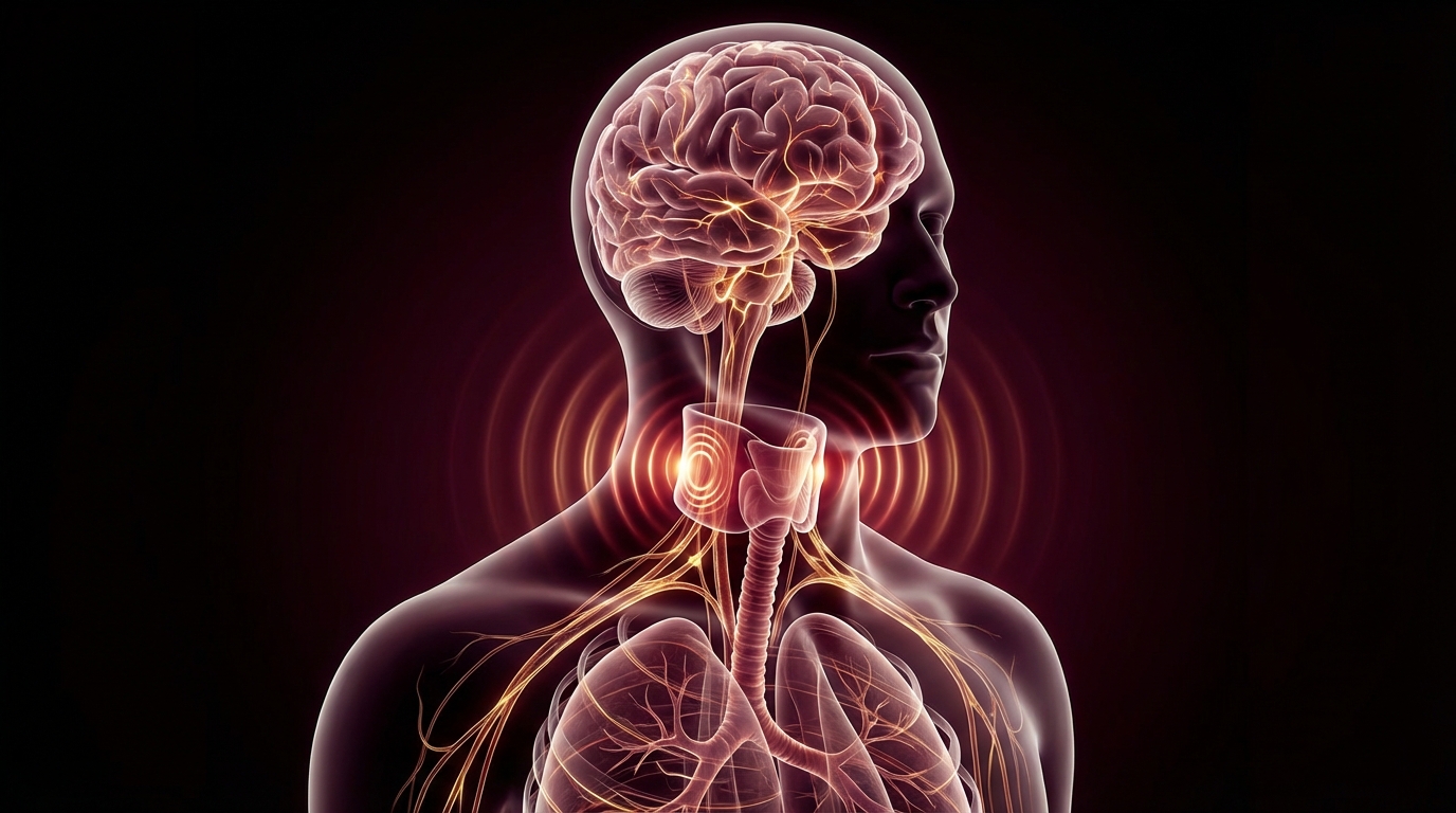
The vagus nerve is your body’s primary brake on inflammation. When it functions properly, it keeps your immune system in check. When it is impaired — as it is in many chronic conditions — inflammation runs unchecked.
In 2000, Kevin Tracey and colleagues at the Feinstein Institute published the finding that made this connection clear. They demonstrated that electrical stimulation of the vagus nerve prevented lethal endotoxic shock in rodents — not by activating a drug or immune pathway previously known, but by triggering a neural reflex that suppressed TNF-alpha production by macrophages [1].
The implication was profound: the nervous system directly controls the immune system’s inflammatory output. Not indirectly through stress hormones. Not vaguely through “mind-body connection.” Directly, through a specific neural circuit with identified neurons, neurotransmitters, and receptors.
This circuit is now called the cholinergic anti-inflammatory pathway, and it may be one of the most clinically relevant discoveries in immunology in the past three decades.
How the Pathway Works
Let me walk through the mechanism step by step, because understanding this pathway changes how you think about inflammation in chronic illness.
Step 1: Inflammatory Signal Detection
When tissue damage or infection occurs, local immune cells (primarily macrophages) release pro-inflammatory cytokines — TNF-alpha, IL-1beta, IL-6. These cytokines signal danger.
Step 2: Afferent Signaling to the Brain
Vagal afferent fibers (sensory fibers running from the body to the brain) detect these inflammatory signals, primarily through cytokine receptors on vagal paraganglia and through the presence of inflammatory mediators in the blood at the blood-brain barrier. This information reaches the nucleus tractus solitarius (NTS) in the brainstem.
Step 3: Central Processing
The brainstem integrates the inflammatory signal and activates vagal efferent (motor) output through the dorsal motor nucleus of the vagus (DMV) and the nucleus ambiguus.
Step 4: The Anti-Inflammatory Reflex
Vagal efferent fibers release acetylcholine at the celiac ganglion, which relays the signal through the splenic nerve to the spleen — the body’s largest reservoir of immune cells. In the spleen, norepinephrine from the splenic nerve activates a specific subset of T cells that release acetylcholine.
Step 5: Macrophage Suppression
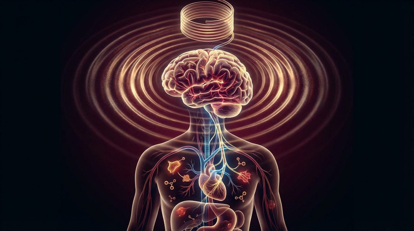
This acetylcholine binds to alpha-7 nicotinic acetylcholine receptors (alpha7nAChR) on macrophages. Activation of these receptors suppresses NF-kB signaling and inhibits the release of TNF-alpha, IL-1beta, IL-6, and HMGB1 — the major drivers of systemic inflammation [2].
The result: the vagus nerve acts as a real-time brake on inflammatory cytokine production. When vagal tone is high (strong vagal activity), inflammation is kept in check. When vagal tone is low (weak vagal activity), the brake is released and inflammation runs unchecked.

Why This Matters for Chronic Illness
Here is where this becomes directly relevant to the patients I treat every day. Chronic inflammatory conditions — Lyme disease, post-COVID syndrome, chronic fatigue, mold illness, autoimmune conditions — share a common feature: persistent, dysregulated inflammation that does not resolve through normal mechanisms.
Multiple lines of evidence now show that impaired vagal tone is both a consequence and a perpetuator of this chronic inflammation:
Lyme Disease
Borrelia burgdorferi infection triggers intense inflammatory responses. But the infection also appears to impair autonomic function directly — many Lyme patients develop dysautonomia with reduced HRV (heart rate variability), a direct measure of vagal tone. Reduced vagal tone means a weakened cholinergic anti-inflammatory pathway, meaning the body’s ability to regulate its own inflammatory response is compromised precisely when it needs that regulation most.
Post-COVID / Long COVID
SARS-CoV-2 has well-documented effects on the autonomic nervous system. Reduced vagal tone has been measured in long COVID patients and correlates with symptom severity. This creates a vicious cycle: the infection triggers inflammation, damages vagal function, which impairs the anti-inflammatory pathway, which allows inflammation to persist, which further damages vagal function.
Chronic Fatigue Syndrome (ME/CFS)
Reduced HRV is one of the most consistent objective findings in ME/CFS research. Multiple studies have documented lower parasympathetic tone and impaired vagal function in CFS patients compared to controls. This provides a mechanistic link between the autonomic dysfunction and the persistent inflammation that characterizes the condition.
POTS (Postural Orthostatic Tachycardia Syndrome)
POTS is fundamentally an autonomic disorder. The vagus nerve is the primary parasympathetic nerve. Impaired vagal function in POTS patients contributes to both the cardiovascular symptoms (inadequate parasympathetic counter-regulation) and the inflammatory symptoms that many POTS patients experience.
The Evidence
What We Know (Human Data)
Vagus nerve stimulation and rheumatoid arthritis: Koopman et al. (2016) published a landmark study in the Proceedings of the National Academy of Sciences showing that implanted vagus nerve stimulation significantly reduced TNF-alpha levels and disease activity in rheumatoid arthritis patients. This was the first human proof-of-concept that the cholinergic anti-inflammatory pathway could be therapeutically targeted [3].
VNS and Crohn’s disease: Bonaz et al. (2016) demonstrated clinical remission in 5 of 7 Crohn’s disease patients treated with vagus nerve stimulation over 12 months. Endoscopic and biological markers of inflammation improved alongside clinical symptoms.
HRV and inflammatory markers: Multiple large epidemiological studies show an inverse correlation between HRV (vagal tone) and circulating inflammatory markers (CRP, IL-6, TNF-alpha). People with higher vagal tone have lower systemic inflammation. This relationship is consistent across healthy populations and disease states.
Non-invasive VNS for long COVID: Preliminary data from multiple centers using transcutaneous vagus nerve stimulation (tVNS) in long COVID patients shows improvements in fatigue, cognitive function, and autonomic markers. A formal clinical trial is evaluating tVNS for long COVID fatigue reduction.
What We See in the Lab (Preclinical)
The preclinical literature is extensive and consistent:
- Vagotomy (cutting the vagus nerve) worsens inflammatory outcomes in virtually every animal model of inflammation
- Vagus nerve stimulation reduces inflammatory markers in models of sepsis, arthritis, colitis, and ischemia-reperfusion injury
- Alpha-7 nicotinic receptor knockout mice show exaggerated inflammatory responses, confirming the receptor’s role
- Acetylcholine and nicotine (as alpha-7 agonists) suppress macrophage TNF-alpha production in cell culture
What I See in Practice
In my clinical experience, patients with the most impaired vagal tone — measured by HRV testing — are consistently the patients with the most difficult-to-control inflammation. This is true across conditions: Lyme, post-COVID, CIRS, autoimmune disease.
What I tell my patients: your body has a built-in anti-inflammatory system, and the vagus nerve is its control wire. When that wire is damaged by infection or illness, inflammation becomes harder to control regardless of what anti-inflammatory treatments we use. Restoring vagal tone is not alternative medicine — it is targeting a well-established neuroimmune pathway that directly controls cytokine production.
Practical Application
How to Assess Your Vagal Tone
The most accessible measure of vagal tone is heart rate variability (HRV). Specifically:
- RMSSD (root mean square of successive differences): A direct measure of parasympathetic (vagal) influence on the heart
- HF power (high-frequency spectral power): Reflects vagal modulation
Consumer wearables (Oura Ring, Whoop, Garmin) provide reasonable HRV estimates. Clinical HRV assessment provides more detailed frequency-domain analysis.
Low HRV in a chronic illness patient suggests impaired vagal tone and a weakened cholinergic anti-inflammatory pathway.
Strategies to Restore Vagal Tone
-
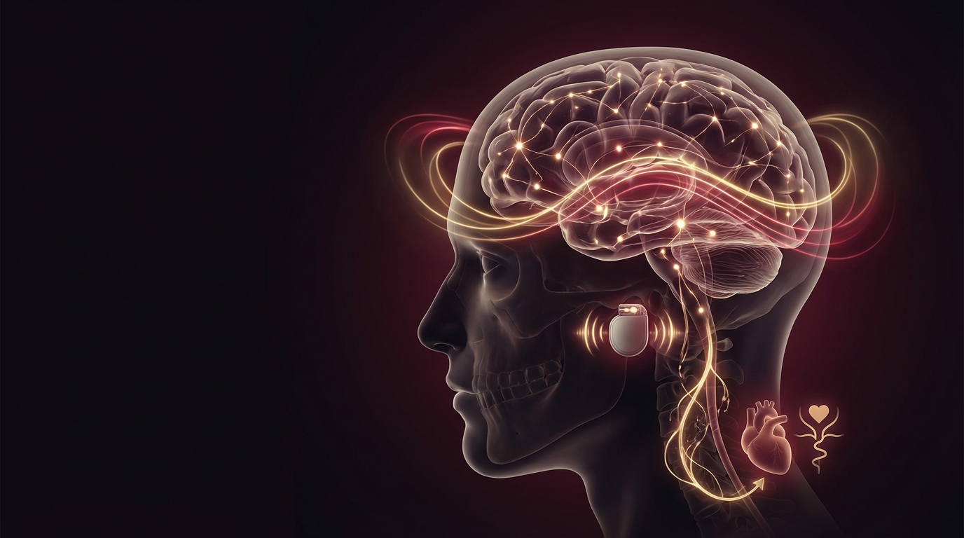
Non-invasive vagus nerve stimulation (tVNS): Devices like gammaCore and Parasym apply electrical stimulation to the auricular branch of the vagus nerve via the ear. This activates the cholinergic anti-inflammatory pathway without surgery.
-
Respiratory-gated HRV biofeedback: Breathing at your resonance frequency (typically 5-7 breaths per minute) maximizes the respiratory sinus arrhythmia and strengthens vagal tone over time. This is the most accessible and evidence-based self-care approach.
-
Cold exposure: Brief cold water exposure (cold shower, face immersion) activates the diving reflex, which is mediated by the vagus nerve. Start with 30 seconds and build gradually. For the full physiological profile of cold water immersion — including how the sympathetic-to-parasympathetic rebound trains vagal resilience — see our cold plunge science guide.
-
Diaphragmatic breathing: Slow, deep belly breathing activates vagal afferents in the diaphragm, stimulating parasympathetic output.
-
Gargling and humming: These activities activate the pharyngeal branch of the vagus nerve. Simple and accessible.
-
Targeted supplementation: Omega-3 fatty acids and probiotics have both been shown to improve HRV in clinical trials, potentially through anti-inflammatory effects on vagal afferent signaling.

Integrating Vagal Tone Restoration into Treatment
For chronic illness patients, vagal tone restoration should be considered alongside — not instead of — treatment of the underlying condition. The sequence I recommend:
- Identify and treat the primary driver (infection, toxin, autoimmune process)
- Assess vagal tone (HRV testing)
- Implement vagal tone restoration strategies
- Monitor HRV and inflammatory markers as treatment progresses
- Consider formal neurofeedback or biofeedback for persistent autonomic dysregulation
Safety and Considerations
Vagus nerve stimulation — both invasive and non-invasive — has a good safety profile. Non-invasive tVNS side effects are minimal: occasional tingling at the stimulation site, rare transient dizziness, and mild cough reflex activation.
Contraindications for tVNS include implanted cardiac devices (pacemaker, defibrillator) and active carotid artery disease. Always consult with your physician before starting any vagus nerve stimulation protocol.
The breathing and lifestyle approaches (HRV biofeedback, cold exposure, diaphragmatic breathing) have essentially no risk when done gradually. Patients with severe dysautonomia should start very conservatively with cold exposure and deep breathing, as these can trigger transient autonomic shifts.
The Bottom Line
The vagus nerve is the body’s primary anti-inflammatory reflex pathway. Through the cholinergic anti-inflammatory mechanism, it directly suppresses macrophage production of TNF-alpha, IL-1beta, and IL-6. When vagal tone is impaired — as it is in many chronic illness patients — inflammation becomes dysregulated and self-perpetuating. Restoring vagal tone is not a wellness trend. It is targeting a specific, well-characterized neuroimmune circuit that controls how much inflammation your body produces. The evidence is clear. The mechanism is established. And for patients stuck in cycles of chronic inflammation, this pathway represents one of the most important therapeutic targets available.
References
- Borovikova LV, Ivanova S, Zhang M, et al. Vagus nerve stimulation attenuates the systemic inflammatory response to endotoxin. Nature. 2000;405(6785):458-462. PMID: 10839541.
- Tracey KJ. The inflammatory reflex. Nature. 2002;420(6917):853-859. PMID: 12490958.
- Koopman FA, et al. Vagus nerve stimulation inhibits cytokine production and attenuates disease severity in rheumatoid arthritis. Proceedings of the National Academy of Sciences. 2016;113(29):8284-8289. PMID: 27382171.
- Bonaz B, et al. Chronic vagus nerve stimulation in Crohn’s disease: a 6-month follow-up pilot study. Neurogastroenterology and Motility. 2016;28(6):948-953. PMID: 26920654.