At a Glance
| Feature | Detail |
|---|---|
| Discovered | 2013 — Maiken Nedergaard, University of Rochester |
| Primary function | Cerebrospinal fluid (CSF) circulation that clears metabolic waste from brain interstitium |
| Peak activity | Slow-wave sleep (N3 stage); up to 10× more active than wakefulness |
| Key waste products cleared | Amyloid-beta, tau, α-synuclein, glutamate, lactate |
| Impaired by | Sleep deprivation, alcohol, sleep apnoea, traumatic brain injury, ageing |
| Clinical relevance | Alzheimer’s prevention, brain fog, neuroinflammation, post-COVID cognition |
| Optimised by | Lateral (side) sleep position, deep sleep architecture, glymphatic-supportive supplements |
Neuroscience spent decades searching for a dedicated lymphatic drainage system in the brain — and declared the brain largely exempt from one. That consensus collapsed in 2013 when Maiken Nedergaard’s laboratory at the University of Rochester published landmark work in Science demonstrating that the brain possesses its own waste-clearance network: the glymphatic system. The name is a portmanteau of glial (the supporting cells that drive it) and lymphatic (the body-wide clearance system it functionally resembles).
What emerged from that discovery and the decade of research that followed reshapes how we think about sleep, neurodegeneration, and cognitive resilience. The glymphatic system is not a passive drain — it is an active, pulsatile circulation that operates on a strict schedule, opening almost exclusively during sleep and shutting down during waking hours. Chronic disruption of that schedule is no longer merely a quality-of-life concern; it is a measurable neurological risk factor.
What the Glymphatic System Is — and How It Works
The brain is enclosed within the skull and therefore cannot swell. Yet it generates enormous metabolic waste: neurons fire thousands of times per second, generating lactate, glutamate, reactive oxygen species, and — critically — the misfolded proteins amyloid-beta and tau that are central to Alzheimer’s pathology.
The body’s conventional lymphatic system does not penetrate brain tissue. Instead, the glymphatic system uses a parallel architecture built from aquaporin-4 (AQP4) water channels on astrocyte endfeet — the same glial cells that surround every blood vessel in the brain.
The mechanism works as follows:
- CSF enters the brain along para-arterial spaces (the space between arterial walls and the surrounding glia)
- Driven by arterial pulsations and, during sleep, by slow electroencephalographic oscillations, CSF is pulled through the brain parenchyma via AQP4 channels
- Interstitial fluid is displaced, carrying metabolic waste products along para-venous channels toward the cervical lymphatics
- Waste is ultimately routed into cervical lymph nodes and from there into systemic circulation for hepatic clearance
The process is elegant and efficient — but only when the system is open. Sleep switches it on. Wakefulness, for reasons still not fully elucidated, suppresses glymphatic flow to roughly 10–15% of its sleep-state capacity.
The Sleep Architecture Connection
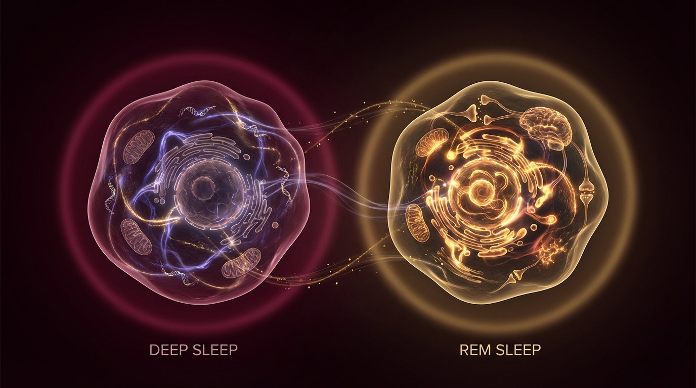
Not all sleep is equal for glymphatic function. Nedergaard’s group and subsequent researchers have consistently shown that slow-wave sleep (SWS, or N3) — characterised by synchronised, high-amplitude delta oscillations below 4 Hz — drives the bulk of glymphatic clearance. These oscillations appear to create pressure differentials that actively pump CSF through peri-vascular channels.
REM sleep contributes to memory consolidation and synaptic plasticity but provides far less glymphatic throughput than SWS. Light sleep (N1/N2) is largely insufficient.
This makes slow-wave sleep depth — not just total sleep duration — a primary target when optimising glymphatic function clinically.
What the Glymphatic System Clears — and Why It Matters
Amyloid-Beta
Amyloid-beta (Aβ) is produced continuously as a byproduct of normal neuronal activity. Under healthy glymphatic conditions, it is cleared nightly. When clearance fails — through sleep disruption, AQP4 dysfunction, or ageing-related glymphatic decline — Aβ accumulates in interstitial spaces, seeds plaques, and triggers the neuroinflammatory cascade that is a hallmark of Alzheimer’s disease.
A 2017 study in Brain demonstrated that a single night of sleep deprivation increased Aβ burden in the human brain by 5% as measured by PET imaging — a sobering demonstration of how rapidly clearance deficits accumulate. Chronic partial sleep restriction (six hours per night for two weeks) produced Aβ elevations comparable to one full night of total sleep deprivation.
Tau
Tau protein stabilises microtubules within neurons. Hyperphosphorylated tau detaches, forms neurofibrillary tangles, and disrupts axonal transport — the second hallmark of Alzheimer’s and primary pathology in frontotemporal dementia (FTD) and chronic traumatic encephalopathy (CTE). Glymphatic clearance of tau follows similar kinetics to Aβ, with nocturnal SWS as the dominant clearance window.
Alpha-Synuclein
α-Synuclein aggregates form Lewy bodies — the cellular signature of Parkinson’s disease and dementia with Lewy bodies (DLB). Glymphatic impairment is now considered a likely contributor to α-synuclein accumulation, with particular relevance to the well-documented link between REM sleep behaviour disorder (RBD) and subsequent Parkinson’s development.
Neuroinflammatory Mediators
Beyond protein aggregates, the glymphatic system clears inflammatory cytokines, excess neurotransmitters, and reactive oxygen species from the brain parenchyma. Impaired clearance creates a low-grade neuroinflammatory milieu that manifests clinically as brain fog, reduced processing speed, and impaired emotional regulation — the same constellation seen in post-COVID-19 neurological sequelae, chronic Lyme disease, and CFS/ME.
Who Is at Highest Risk of Glymphatic Dysfunction?
Several clinical populations warrant focused attention on glymphatic health:
Chronic poor sleepers: Even mild but consistent sleep restriction (habitually sleeping six hours or fewer) produces measurable glymphatic impairment. The data suggest that cumulative “sleep debt” cannot be fully repaid by weekend recovery sleep — the nightly clearance opportunity, once missed, is gone.
Sleep apnoea patients: Obstructive sleep apnoea fragments slow-wave sleep and creates intermittent hypoxia. Multiple studies confirm elevated Aβ and tau burden in untreated OSA patients; CPAP therapy partially — but not fully — reverses this.
Post-COVID and long-COVID patients: Emerging evidence implicates glymphatic dysfunction in post-COVID neurological symptoms. Microglial activation, astrocyte AQP4 redistribution, and disrupted sleep architecture all converge to impair clearance. This may explain why cognitive symptoms persist months after viral clearance.
Traumatic brain injury (TBI): TBI disrupts AQP4 channel polarity on astrocyte endfeet — the molecular machinery that drives glymphatic flow. This likely contributes to the elevated dementia risk observed in post-TBI populations, including athletes with repeated concussion.
Ageing adults: Glymphatic efficiency declines approximately 0.5–1% per year after age 40, correlated with declining SWS duration, arterial stiffening (which reduces the pulsatile driving force), and AQP4 redistribution. This makes glymphatic optimisation particularly relevant in the longevity context.
Chronic alcohol users: Alcohol suppresses SWS and redistributes AQP4 channels. Even moderate alcohol consumption in the hours before sleep measurably reduces glymphatic throughput the following night.
How to Optimise Glymphatic Function Clinically
1. Prioritise Deep Sleep Architecture
The primary intervention is maximising SWS. Practical targets include:
- Sleep duration: 7–9 hours for adults; each hour reduction below 7 hours imposes measurable glymphatic cost
- Sleep timing consistency: Circadian alignment maximises SWS allocation in the first sleep cycle (typically 11 pm–1 am)
- Blue light restriction: Melatonin suppression from blue light shifts sleep onset and compresses early SWS
- Alcohol avoidance within 3 hours of sleep: Even two standard drinks disrupt SWS by 20–30%
2. Lateral Sleep Position
One of the more surprising findings in glymphatic research is that sleep position substantially affects clearance efficiency. A 2019 rodent study (and corroborated by human imaging data) showed that lateral (side-lying) sleep — particularly right lateral — produces superior CSF-ISF exchange compared with supine or prone positioning. This is attributed to gravity-assisted peri-vascular flow and better airway patency reducing arousal frequency.
Right lateral positioning consistently outperforms left lateral in glymphatic studies, possibly due to cardiac position effects on cerebrovascular pulsatility.
3. Supplement-Based Glymphatic Support
Several compounds have mechanistic evidence for supporting glymphatic function:
Magnesium L-threonate (145–200 mg elemental, evening): Crosses the blood-brain barrier more efficiently than other magnesium forms; increases synaptic density and has demonstrated SWS-enhancing effects in human trials.
Lion’s mane mushroom (Hericium erinaceus, 500–1000 mg daily): Stimulates nerve growth factor (NGF) synthesis and appears to support AQP4 expression and astrocyte health. Particularly relevant for post-COVID and early cognitive decline.
Low-dose melatonin (0.3–1.0 mg, 60–90 minutes pre-sleep): Physiological-range dosing (not the 5–10 mg “sleep-aid” doses) more accurately replicates endogenous melatonin kinetics and avoids receptor downregulation. Melatonin has direct anti-amyloid effects independent of its sleep-promoting action.
Glycine (3 g, pre-sleep): A major inhibitory neurotransmitter with demonstrated SWS-deepening effects in human RCTs; reduces core body temperature, which is a key driver of SWS initiation.
Omega-3 fatty acids (EPA+DHA, 2–4 g daily): Modulate neuroinflammation and maintain astrocyte membrane fluidity critical for AQP4 channel function.
4. Address Upstream Sleep Disorders
Sleep apnoea, restless legs syndrome, and circadian rhythm disorders directly fragment SWS. CPAP therapy for OSA improves — though does not fully normalise — glymphatic clearance markers. Treating RLS with magnesium, iron optimisation, or dopaminergic agents preserves sleep architecture.
5. Neurofeedback for SWS Enhancement
Closed-loop neurofeedback protocols targeting delta oscillation amplitude have shown promise in enhancing SWS depth in adults with disrupted sleep architecture. This is an area of active clinical interest, particularly for patients with post-COVID cognition or CFS/ME where pharmacological sleep interventions carry risks.
6. Cold and Heat Exposure Timing
Morning cold exposure (3–5 minutes cold shower or plunge) raises noradrenaline and improves circadian entrainment, indirectly supporting SWS allocation. Sauna use 2–3 hours before sleep — not immediately before — raises then lowers core body temperature, mimicking the temperature drop that cues SWS onset. Sauna immediately before bed suppresses the temperature signal and can paradoxically reduce SWS.
The Clinical Picture: Brain Fog, Neurodegeneration, and the Sleep-Immunity Connection
In clinical practice, glymphatic dysfunction rarely presents as an isolated finding. It is embedded in a network of bidirectional relationships:
Neuroinflammation impairs sleep → impaired sleep worsens neuroinflammation. Cytokines including IL-1β, TNF-α, and IL-6 disrupt sleep architecture, reducing SWS. Reduced SWS means reduced clearance of these same mediators. This creates the self-reinforcing cycle seen in post-infectious cognitive syndromes.
Autonomic dysfunction affects CSF pulsatility. The driving force for glymphatic flow is arterial pulsation transmitted through peri-vascular channels. In conditions characterised by autonomic dysregulation — post-COVID dysautonomia, POTS, chronic Lyme disease — blunted arterial pulsatility may reduce the mechanical driving force for CSF circulation.
HPA axis dysregulation and evening cortisol. Elevated evening cortisol — a finding in chronic stress, adrenal fatigue states, and HPA axis dysregulation — directly suppresses SWS onset and duration. Addressing cortisol rhythmicity (via adaptogen protocols, circadian light exposure, and stress reduction) is therefore a relevant upstream glymphatic intervention.
When patients present with persistent brain fog that does not resolve with anti-inflammatory treatment alone, the question of glymphatic efficiency deserves explicit clinical attention. Sleep architecture analysis (actigraphy or polysomnography) belongs in the workup alongside inflammatory panels and autonomic assessment.
Related Articles
- Deep Sleep: How to Increase Slow-Wave Sleep Naturally
- Magnesium and Sleep: Which Form Works Best?
- Lion’s Mane Mushroom: Evidence for Nerve Regeneration and Cognitive Protection
- Post-COVID Brain Fog: Mechanisms, Testing, and Treatment
- Neurofeedback for ADHD and Cognitive Enhancement
References
-
Iliff JJ, Wang M, Liao Y, et al. A paravascular pathway facilitates CSF flow through the brain parenchyma and the clearance of interstitial solutes, including amyloid β. Sci Transl Med. 2012;4(147):147ra111. PMID: 22896675
-
Xie L, Kang H, Xu Q, et al. Sleep drives metabolite clearance from the adult brain. Science. 2013;342(6156):373–377. PMID: 24136970
-
Shokri-Kojori E, Wang GJ, Wiers CE, et al. β-Amyloid accumulation in the human brain after one night of sleep deprivation. Proc Natl Acad Sci USA. 2018;115(17):4483–4488. PMID: 29632177
-
Lee H, Xie L, Yu M, et al. The effect of body posture on brain glymphatic transport. J Neurosci. 2015;35(31):11034–11044. PMID: 26245965
-
Benveniste H, Liu X, Koundal S, et al. The glymphatic system and waste clearance with brain aging: a review. Gerontology. 2019;65(2):106–119. PMID: 29996134
-
Holth JK, Fritschi SK, Wang C, et al. The sleep-wake cycle regulates brain interstitial fluid tau in mice and CSF tau in humans. Science. 2019;363(6429):880–884. PMID: 30679382
-
Ding F, O’Donnell J, Xu Q, et al. Changes in the composition of brain interstitial ions control the sleep-wake cycle. Science. 2016;352(6285):550–555. PMID: 27126038