If you are in acute danger, contact emergency services immediately or call your national crisis line.
At a Glance
| Property | Detail |
|---|---|
| Key biomarker | High-sensitivity CRP (hs-CRP) |
| Inflamed subtype prevalence | ~27% of patients with major depressive disorder |
| TRD association | CRP significantly elevated in treatment-resistant vs. treatment-responsive depression after BMI correction |
| TMS implication | Elevated CRP associated with attenuated rTMS response; cytokine reductions during TMS track antidepressant effect |
| CRP threshold for action | hs-CRP ≥ 3 mg/L warrants investigation before proceeding |
| Priority | Rule out occult infections before attributing high CRP to lifestyle alone |
| Testing sequence | hs-CRP first; full cytokine panel if elevated; infection screen if CRP > 3 mg/L |
The question is not whether you should start TMS. The question is whether TMS, started today, will actually work — or whether you are about to run a brain stimulation protocol against a background of systemic inflammation that will blunt every session.
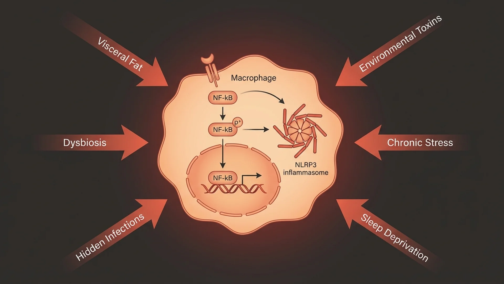
About one in four patients with major depressive disorder has detectable systemic inflammation, most readily measured by peripheral high-sensitivity C-reactive protein. For those patients, the standard antidepressant pathway — and the standard TMS referral — consistently underdelivers, not because TMS doesn’t work, but because inflammation is actively interfering with the neurochemical substrates that make it work. The correct sequence is not to try TMS and hope for the best. It is to test first, treat the inflammation if needed, and only then apply brain stimulation to a system that can respond.
This is not a theoretical concern. It is what the data show, and it is what clinical experience confirms.
Who Has the Inflamed Subtype — and What It Looks Like
Estimates of how often neuroinflammation contributes to depression cluster around 27% of patients with major depressive disorder (Hassamal, 2023, PMID 37252156). That is roughly one in four — a large enough minority that inflammation testing is not optional in any thorough workup. In treatment-resistant depression, the proportion is higher.
The critical study here is Chamberlain et al. (2019, PMID 29764522), published in the British Journal of Psychiatry. It compared CRP levels in four groups: 102 patients with treatment-resistant MDD, 48 with treatment-responsive MDD, 48 with untreated MDD, and 54 healthy volunteers. After correcting for BMI — the most important confounder — CRP was significantly elevated only in the treatment-resistant group (Cohen’s d = 0.47). The treatment-responsive and untreated groups were not significantly different from healthy controls.
This matters enormously. It means that CRP elevation is not just a correlate of depression — it is a correlate of failed treatment. Patients who are not getting better are systematically more inflamed than patients who are.
Clinically, the inflamed subtype does not always look like the textbook depressed patient. Fellows et al. (2024, PMID 39359158), studying 168 patients with treatment-resistant depression in a cross-cultural cohort, found that high CRP (≥ 3 mg/L) was specifically associated with early-night insomnia, early-morning waking, and psychic anxiety. Cognitive symptoms — the ones that often remain even after mood partially improves — may also be more persistent in patients with elevated inflammatory markers. When a patient describes their depression as primarily driven by anxious agitation, sleep-onset and sleep-maintenance problems, and a sense that something biological is wrong, inflammation belongs on the differential.
The Testing Panel: What to Order and What the Numbers Mean
High-sensitivity CRP is the first-line test, for the same reason it has entered cardiovascular medicine: it is inexpensive, widely available, stable under standard laboratory handling, and peripheral CRP levels correlate closely with cerebrospinal fluid CRP (Hassamal, 2023). Ordering the standard CRP is insufficient for this purpose — only the hs-CRP assay has the sensitivity to detect the low-grade elevations that are clinically meaningful in depression.
hs-CRP thresholds in clinical context:
| hs-CRP level | Interpretation | Clinical implication |
|---|---|---|
| < 1.0 mg/L | Low-risk | Inflammation unlikely to be a primary driver |
| 1.0–3.0 mg/L | Moderate elevation | Low-grade inflammation; lifestyle factors likely; test for occult causes if TRD |
| 3.0–5.0 mg/L | High | Inflammation is a probable driver; investigate before TMS |
| > 5.0 mg/L | Elevated (“hot”) | Anti-inflammatory intervention warranted; infliximab trial data apply; urgent infection/autoimmune screen |
Beyond hs-CRP, the full inflammatory picture adds meaningful resolution. A practical first-tier extension panel includes:
- Interleukin-6 (IL-6): Crosses the blood-brain barrier and directly suppresses serotonin synthesis. Elevated IL-6 also downregulates hippocampal neurogenesis, which is one of the postulated mechanisms of TMS’s antidepressant effect.
- TNF-alpha: Wang et al. (2022, PMID 35722555) demonstrated in a 57-patient prospective sham-controlled trial that early drops in TNF-alpha during rTMS predicted clinical improvement at week 2. TNF-alpha at baseline therefore carries prognostic weight.
- Neutrophil-to-lymphocyte ratio (NLR): An accessible whole-blood calculation that tracks systemic immune activation. Emerging data show that elevated pre-treatment NLR is associated with attenuated rTMS response in treatment-resistant depression.
- Ferritin: Elevated ferritin is both a marker of acute-phase inflammation and, separately, a marker of iron overload. It is worth drawing when CRP is elevated to distinguish inflammatory ferritin from primary iron excess.
- Erythrocyte sedimentation rate (ESR): Slower to change than CRP and less specific, but adds value when temporal trends are needed or when vasculitis or autoimmune disease is suspected.
How Elevated CRP Undermines Antidepressant and TMS Response
The mechanism is not a black box. Inflammation disrupts depression treatment through several converging pathways:
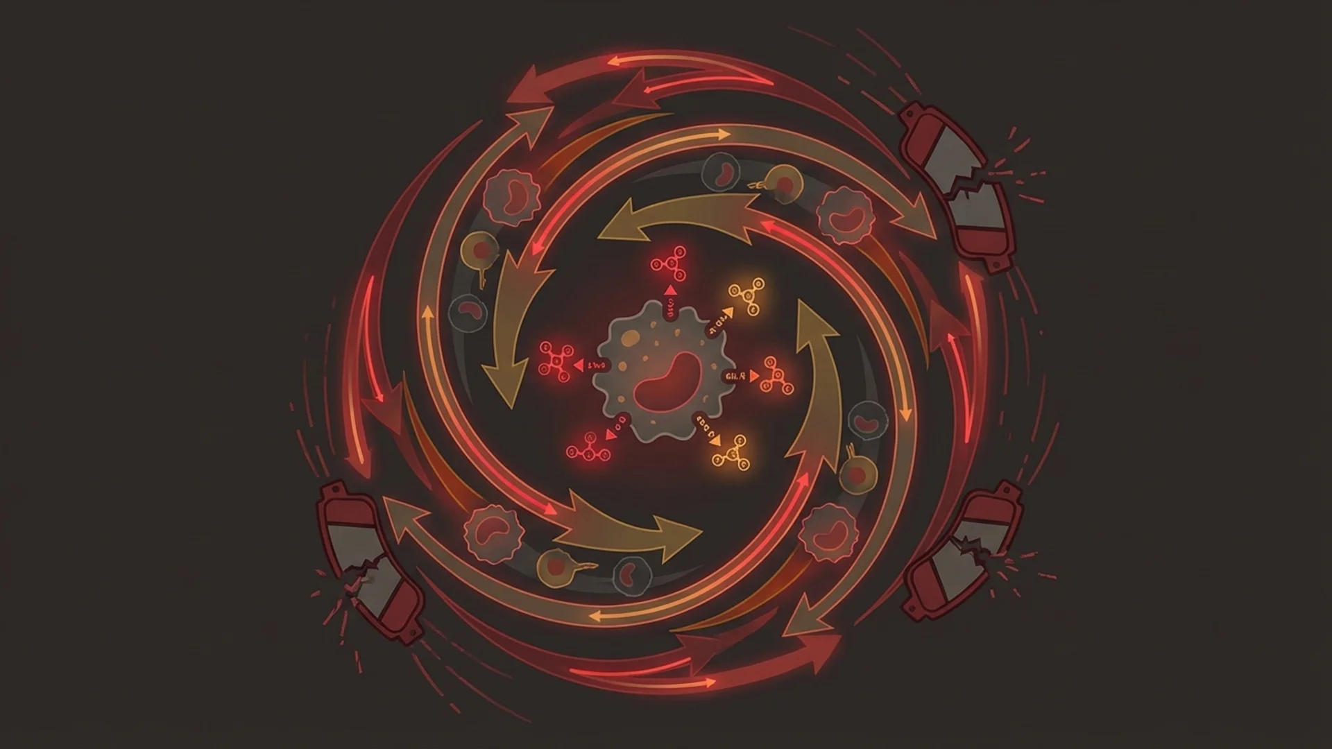
The IDO pathway. Elevated pro-inflammatory cytokines — particularly IL-6, TNF-alpha, and IFN-gamma — activate the enzyme indoleamine-2,3-dioxygenase (IDO), which shunts the metabolism of tryptophan away from serotonin and toward the kynurenine pathway. The result is reduced serotonin availability in the prefrontal cortex — the same region targeted by rTMS for depression. TMS applied to a serotonin-depleted left dorsolateral prefrontal cortex is working against biology.
BDNF suppression. Inflammatory cytokines reduce brain-derived neurotrophic factor expression. BDNF is central to the plasticity mechanisms through which rTMS is thought to consolidate antidepressant effects. Low BDNF, sustained by ongoing inflammation, limits how much structural change TMS can induce.
Direct meta-analytic evidence. Gasparini et al. (2022, PMID 35342200) conducted a systematic review and meta-analysis of 31 studies examining inflammatory biomarkers and antidepressant response in MDD. In the CRP-specific analysis (seven studies, 1,493 patients), baseline CRP was significantly higher in non-responders compared to responders (effect size −0.18, 95% CI −0.28 to −0.08; p = 0.0006, I² = 0%). Zero heterogeneity across studies is unusual and strengthens the signal. IL-8 was also significantly higher in non-responders.
At the extreme end. Raison et al. (2013, PMID 22945416) tested infliximab, a TNF-alpha blocker, against placebo in treatment-resistant depression. Across the full sample, infliximab had no advantage. But in the subgroup with baseline hs-CRP above 5 mg/L — the actively inflamed patients — response rates were 62% with infliximab versus 33% with placebo. This is an exploratory finding from a small sample, but its implication is clear: patients with high baseline CRP appear to be in a biologically distinct category, one where the immune system is driving the depression and must be addressed directly.
For TMS specifically, the Wang et al. trial (2022, PMID 35722555) showed that in patients receiving active rTMS, changes in IL-2 and high-sensitivity CRP tracked clinical improvement across the treatment course. The patients who improved showed greater inflammatory marker reductions. Whether this relationship is causal — whether reducing inflammation before TMS would amplify the response — is not yet settled in a prospective trial, but the mechanistic case is strong enough to warrant the precaution.
The Decision Tree: What to Do with the CRP Result
hs-CRP < 1.0 mg/L: Inflammation is unlikely to be a meaningful driver. Proceed with the standard workup for depression (qEEG brain mapping, infection screen if clinically indicated) and initiate TMS according to the network pattern identified on mapping.
hs-CRP 1.0–3.0 mg/L: Low-grade chronic inflammation. Address lifestyle factors (sleep, exercise, diet, stress) alongside TMS rather than waiting. This level seldom requires a prolonged delay before TMS but should be followed up at the end of the stimulation course.
hs-CRP 3.0–5.0 mg/L: Clinically meaningful elevation. Before TMS, complete a targeted cause investigation: Lyme disease and co-infections (Bartonella, Babesia, Ehrlichia), reactivated viruses (EBV, HHV-6), autoimmune screening (ANA, anti-dsDNA, thyroid antibodies), and gut integrity markers (zonulin, calprotectin). If an infectious or autoimmune cause is found, treat it first. If no discrete cause is identified, implement a structured eight-to-twelve-week anti-inflammatory protocol before beginning TMS. Retest hs-CRP before the first session.
hs-CRP > 5.0 mg/L: A mandatory infection and autoimmune workup before any further treatment decisions. This level corresponds to the threshold identified by Raison et al. (2013) where anti-cytokine therapy showed signal. At this level, inflammation is not a bystander — it is driving the clinical picture. Targeted TMS into this environment has a low probability of full response and may waste a treatment course that the patient cannot easily repeat.
Treating Inflammation Before TMS: The Protocol
The goal is to bring hs-CRP below 3 mg/L — ideally below 1 mg/L — before beginning brain stimulation. For most patients with idiopathic low-grade inflammation (no discrete infection or autoimmune driver), an eight-to-twelve-week anti-inflammatory protocol achieves this.
Step 1 — Rule out infections first. This cannot be skipped. Lyme disease, Bartonella, reactivated EBV, and HHV-6 are under-recognized causes of persistent high CRP and neuropsychiatric symptoms that look identical to treatment-resistant depression. Standard immunoassays miss chronic Lyme frequently; a clinical specialist with experience in tick-borne illness and atypical presentations should be part of the workup when CRP stays high and standard causes are absent.
Step 2 — Dietary foundation. A Mediterranean-pattern diet consistently reduces hs-CRP in meta-analyses, with effect sizes larger than most pharmaceutical interventions studied for inflammation. Eliminating ultra-processed foods, refined carbohydrate, and seed oils removes the most potent inflammatory substrates. This is non-negotiable, not optional.
Step 3 — Targeted supplementation. Omega-3 fatty acids (EPA + DHA, minimum 2–3 grams per day of combined EPA and DHA) are the highest-evidence anti-inflammatory supplement. Curcumin with piperine, berberine, and high-dose vitamin D3 (with K2) add sequential benefit. These are not substitutes for identifying and removing the inflammatory cause.
Step 4 — Sleep and exercise. Both independently reduce IL-6 and CRP. Seven to nine hours of consolidated sleep is not a lifestyle preference in this context — it is part of the treatment protocol.
Step 5 — Retest and decide. After eight to twelve weeks, recheck hs-CRP and the extended cytokine panel. If the level has normalized, proceed to brain mapping and TMS with an expectation of full response capacity. If the level remains elevated despite the protocol, escalate the infection/autoimmune investigation before proceeding.
Related Articles
- Treatment-Resistant Depression: The Cause-First Workup Before TMS — The full four-domain protocol: brain networks, inflammation, infections, and relationships.
- TMS for Depression: What 20 Years of Evidence Shows — The clinical evidence base for rTMS in depression and how to interpret response rates.
- Chronic Inflammation: The Root of Disease — Mechanisms of systemic low-grade inflammation and evidence-based strategies for reducing it.
- Brain Fog and Neuroinflammation: The PET Scan Evidence — Microglial activation and central nervous system inflammation documented in Lyme and post-infectious states.
- ECT Side Effects and Alternatives — Why ECT should not be the escalation step for treatment-resistant depression, and what the evidence shows.
References
-
Chamberlain SR, Cavanagh J, de Boer P, et al. Treatment-resistant depression and peripheral C-reactive protein. Br J Psychiatry. 2019;214(1):11–19. doi:10.1192/bjp.2018.66. PMID: 29764522
-
Gasparini A, Callegari C, Lucca G, et al. Inflammatory Biomarker and Response to Antidepressant in Major Depressive Disorder: a Systematic Review and Meta-Analysis. Psychopharmacol Bull. 2022;52(1):36–52. doi:10.64719/pb.4425. PMID: 35342200
-
Wang Q, Zeng L, Hong W, et al. Inflammatory Cytokines Changed in Patients With Depression Before and After Repetitive Transcranial Magnetic Stimulation Treatment. Front Psychiatry. 2022;13:925007. doi:10.3389/fpsyt.2022.925007. PMID: 35722555
-
Raison CL, Rutherford RE, Woolwine BJ, et al. A Randomized Controlled Trial of the Tumor Necrosis Factor-alpha Antagonist Infliximab in Treatment Resistant Depression: Role of Baseline Inflammatory Biomarkers. JAMA Psychiatry. 2013;70(1):31–41. doi:10.1001/2013.jamapsychiatry.4. PMID: 22945416
-
Fellows E, Jones BDM, Hodsoll J, et al. Associations between C-reactive protein and individual symptoms of depression in a lower-middle income country. BJPsych Open. 2024;10(5):e169. doi:10.1192/bjo.2024.735. PMID: 39359158
-
Hassamal S. Chronic stress, neuroinflammation, and depression: an overview of pathophysiological mechanisms and emerging anti-inflammatories. Front Psychiatry. 2023;14:1130989. doi:10.3389/fpsyt.2023.1130989. PMID: 37252156