At a Glance
| Feature | Detail |
|---|---|
| Condition | Postural Orthostatic Tachycardia Syndrome (POTS) / Dysautonomia |
| Core mechanism | Failure of autonomic cardiovascular regulation on standing |
| Diagnostic criterion | HR increase ≥ 30 bpm within 10 min of standing (≥ 40 bpm in under-19s) |
| Common triggers | Post-viral infection, Lyme disease, autoimmunity, trauma, surgery |
| Functional medicine targets | Neuroinflammation, volume depletion, mitochondrial dysfunction, MCAS, vagal tone |
| Key tests | Active Stand Test, TTT, autoimmune panels, catecholamines, tryptase, lactulose breath test |
| Expected timeline | 6–18 months for meaningful improvement with comprehensive protocol |
Postural Orthostatic Tachycardia Syndrome was once considered rare. It is no longer. Conservative estimates place prevalence at 1–3 million in the US alone, and neurologists are now describing what epidemiologists are calling a secondary pandemic: post-infectious dysautonomia driven primarily by SARS-CoV-2 but also by Lyme disease, Epstein–Barr virus reactivation, and other immune-activating infections.
Patients arrive at my clinic after years of being told their resting ECG looks fine. They have been prescribed beta-blockers, told to drink more water, and sent home with compression garments. Some improve slightly. Most do not — because the mechanism driving their POTS has not been identified, let alone treated.
Conventional cardiology manages the symptom: the heart rate spike on standing. Functional medicine asks a different question: why has the autonomic nervous system lost the ability to regulate cardiovascular tone in the first place? The answer to that question — and it is almost always a multi-layered one — determines the protocol.
What Is POTS? The Autonomic Physiology You Need to Understand
The autonomic nervous system (ANS) continuously adjusts vascular tone, heart rate, and blood volume distribution in response to gravitational challenge. When you stand, approximately 500–700 mL of blood pools in the lower extremities. Healthy baroreceptors detect this, activate sympathetic pathways, and cause peripheral vasoconstriction and a modest 10–15 bpm heart rate increase within seconds.
In POTS, this reflex is broken. The compensatory vasoconstriction does not occur adequately, blood continues to pool, cerebral perfusion drops, and the heart rate surges — often 30–60 bpm above baseline — in a desperate attempt to maintain cardiac output. The patient feels dizzy, cognitively impaired, breathless, and exhausted.
The Subtypes That Change Your Treatment
POTS is not one disease. Conflating the subtypes is the primary reason treatment protocols fail:
- Hypovolemic POTS: low blood volume (low renin, low aldosterone); responds to volume expansion and fludrocortisone
- Hyperadrenergic POTS: excess standing norepinephrine (>600 pg/mL); responds to central sympatholytic agents; beta-blockers can worsen it
- Neuropathic POTS: small fiber neuropathy reducing peripheral vasoconstriction; confirmed by skin punch biopsy
- Post-infectious / autoimmune POTS: adrenergic receptor autoantibodies or anti-ganglionic AChR antibodies; the fastest-growing subtype
- Mast cell–associated POTS: MCAS-driven histamine and prostaglandin release; flushing, GI symptoms, and urticaria are clues
Identifying the dominant subtype — through catecholamine fractionation, autoantibody testing, and a detailed symptom map — is not optional. It is the foundation of a rational protocol.
Root Causes: The Functional Medicine Diagnostic Framework
Neuroinflammation and Post-Viral Damage
The link between viral infection and POTS is now mechanistically established. SARS-CoV-2 spike protein has been detected in autonomic ganglia at autopsy. Molecular mimicry between viral antigens and adrenergic or muscarinic receptors triggers autoantibody production. Neuroinflammatory cytokines — particularly IL-6, TNF-α, and interferon-gamma — damage small fiber neurons that carry autonomic traffic.
In our post-COVID patient cohort, we routinely measure adrenergic receptor autoantibodies (β1, β2, α1, muscarinic M2/M3). Elevated titres — found in approximately 40–60% of post-COVID POTS patients in published series — guide immunomodulatory treatment decisions.
Small Fiber Neuropathy
Small unmyelinated C-fibers and lightly myelinated Aδ-fibers carry autonomic vasoconstrictor signals to peripheral vessels. When these are damaged — by infection, autoimmunity, metabolic disease, or toxin exposure — the vessels in the lower extremities cannot constrict on command.
Skin punch biopsy at the distal leg (measuring intraepidermal nerve fiber density) is the diagnostic standard. Low fiber density (below the 5th percentile for age and sex) confirms neuropathic POTS and justifies aggressive neuroprotective protocols including IVIG in selected cases.
Mitochondrial Dysfunction
ATP production is the engine of every autonomic reflex. Mitochondrial dysfunction — accelerated by oxidative stress, viral infection, and persistent neuroinflammation — depletes the energy reserves that autonomic neurons and cardiac pacemaker cells require for high-frequency firing.
Clinically, this presents as post-exertional malaise that follows the same curve as ME/CFS: a functional plateau followed by crash, not a simple linear fatigue. Organic acid testing, CoQ10 levels, and carnitine fractionation map this component. Supplementation alone rarely reverses it; we use IV NAD⁺, mitochondria-targeted antioxidants, and structured low-intensity aerobic reconditioning (horizontal exercise initially) as a combined approach.
Volume and Hormonal Dysregulation
Low blood volume is both a driver and a consequence of POTS. Reduced gravitational stress from bedrest, ANS-mediated changes in renal handling, and suppressed aldosterone all contribute. We measure plasma renin activity and aldosterone along with a 24-hour urine sodium to guide volume replacement.
Many patients — particularly women in the luteal phase — also show progesterone-driven venous pooling. Hormonal profiling is not optional in female patients with cycle-linked POTS exacerbations.
Diagnostic Workup: Beyond the Tilt Table Test
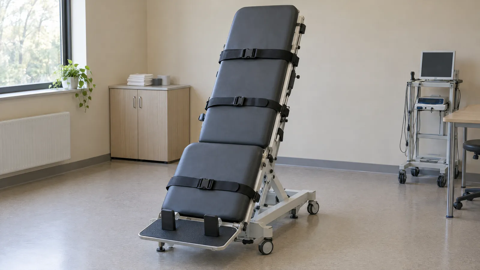
The tilt table test (TTT) confirms POTS. It does not explain it. Our diagnostic battery adds the following:
Autoimmune and inflammatory:
- Adrenergic receptor autoantibodies (β1, β2, α1, M2, M3)
- Anti-ganglionic AChR antibodies
- ANA, anti-dsDNA, anti-Ro/La, anti-CCP
- Cytokine panel (IL-6, IL-8, TNF-α, TGF-β)
- Tryptase and 24-hour urine prostaglandin D2 (for MCAS)
Autonomic and neurological:
- Quantitative Sudomotor Axon Reflex Test (QSART) where available
- Skin punch biopsy (distal leg intraepidermal nerve fiber density)
- Thermoregulatory sweat test
- Plasma catecholamines supine and standing (10 min)
Volume and hormonal:
- Plasma renin activity and aldosterone (supine and standing)
- 24-hour urine sodium, potassium, catecholamines
- Full hormonal panel: cortisol (4-point salivary), DHEA-S, sex hormones
Infectious triggers:
- Comprehensive Lyme and co-infection panel (iSpot Lyme, Bartonella FISH, EBV/CMV/HHV-6 serology with avidity)
- COVID spike protein persistence assay where available
The Functional Medicine Protocol for POTS
Volume Expansion and Electrolyte Optimization
High sodium intake (5–10 g/day from food and supplementation) combined with aggressive hydration (2.5–3 L/day) is the first-line intervention for hypovolemic POTS. Electrolyte drinks with sodium, potassium, and magnesium are preferable to plain water, which can worsen hyponatremia and trigger reflex vasodilation.
In patients with confirmed low renin and aldosterone, pharmaceutical fludrocortisone (0.05–0.2 mg/day) is a reasonable short-term bridge while root causes are addressed. We do not use it as a long-term substitute for identifying why volume regulation is impaired.
IV saline infusions (1–2 L normal saline) can provide dramatic short-term relief for severe exacerbations — the effect typically lasts 24–48 hours and confirms the hypovolemic component. Some patients maintain a schedule of weekly IV saline during the recovery phase.
Mitochondrial Support Protocol
- CoQ10 (ubiquinol form): 200–400 mg/day: addresses electron transport chain dysfunction common in post-viral states
- NAD⁺ precursors: NMN 500 mg or NR 1,000 mg daily; IV NAD⁺ (500–1,000 mg infusions) for more severe presentations
- L-carnitine / acetyl-L-carnitine: 1,500–2,000 mg/day for fatty acid transport support
- Magnesium glycinate: 400–600 mg/day: cofactor for over 300 enzymatic reactions including ATP synthesis
- Alpha-lipoic acid (R-form): 300–600 mg/day: mitochondrial antioxidant with documented benefit in small fiber neuropathy
Mast Cell Stabilization
When MCAS is confirmed or strongly suspected (elevated tryptase, prostaglandins, or histamine; symptom pattern of flushing, GI cramps, and urticaria with positional changes), mast cell stabilization is a prerequisite, not an adjunct. Histamine and prostaglandin release from activated mast cells directly cause vasodilation and worsen pooling.
Protocol: second-generation H1 blocker (cetirizine or loratadine, 10–20 mg twice daily) combined with H2 blocker (famotidine, 20–40 mg twice daily), plus quercetin (500–1,000 mg twice daily as a natural mast cell stabilizer) and vitamin C (2–3 g/day). Cromolyn sodium (oral) is added for patients with predominant GI involvement.
Dietary modification — low-histamine diet for 4–6 weeks while the protocol stabilizes — reduces the inflammatory substrate.
Vagus Nerve Rehabilitation
The vagus nerve is the primary parasympathetic brake on heart rate. Reduced vagal tone — measurable via heart rate variability (HRV) monitoring — is a consistent finding in POTS and a driver of persistent sympathetic dominance.
Vagus nerve rehabilitation is graduated:
- Breathing exercises: 4-7-8 or resonance frequency breathing (5.5 breaths/min) for 20 minutes daily — documented to increase HRV in multiple trials
- Cold water face immersion: The diving reflex powerfully activates vagal pathways; 30-second cold water face immersion lowers heart rate by 10–20 bpm in most patients
- Non-invasive vagus nerve stimulation (nVNS): Transcutaneous auricular VNS devices are now available OTC; clinical evidence supports HRV improvement with 15–30 minutes daily use
- Horizontal aerobic reconditioning: The exercise intolerance in POTS is real but not a reason to avoid all movement. Rowing machines, recumbent bikes, and swimming allow cardiovascular conditioning without the gravitational challenge of upright exercise. Progressive increase over 3–6 months rebuilds cardiac chamber volume and autonomic calibration.
Post-COVID POTS: A Special Case
Post-COVID POTS differs from classic POTS in several ways that alter protocol design. The autoimmune burden is higher, spike protein persistence in tissues may drive ongoing inflammation, and micro-clot formation (documented in multiple post-COVID autopsy series) can compromise microvascular perfusion in autonomic ganglia.
In our post-COVID POTS patients, we add:
- Low-dose naltrexone (LDN): 1.5–4.5 mg at night: modulates microglial activation and pro-inflammatory cytokines; well-tolerated with minimal side-effect profile
- Nattokinase and lumbrokinase: For documented microclot burden; fibrinolytic enzymes address the vascular component
- Apheresis consideration: In patients with elevated autoantibodies and poor response to standard measures, therapeutic apheresis (HELP or immunoadsorption) can remove circulating autoantibodies and inflammatory mediators; we have seen rapid improvement in heart rate variability and orthostatic tolerance following apheresis in this subgroup
What to Expect: Realistic Outcomes and Timeline
POTS is not a life sentence. In our clinic, patients with clear post-infectious POTS and an identified dominant mechanism achieve meaningful functional improvement in 6–12 months with comprehensive treatment. Those with long-standing neuropathic damage or high autoantibody titres require 12–18 months.
The sequence matters. We address volume and electrolytes first (days to weeks for response), then mast cell stabilization if indicated (weeks), then mitochondrial support and vagal rehabilitation (months), then upstream immunological drivers. Trying to rehabilitate exercise capacity before stabilizing volume is a common error that leads to post-exertional crashes and loss of patient confidence.
Objective monitoring via HRV tracking (a smartwatch is sufficient) gives both patient and physician a longitudinal window into autonomic recovery that symptom questionnaires alone cannot provide.
Related Articles
- Post-COVID Brain Fog: Neuroinflammation and Recovery Strategies
- Vagus Nerve Stimulation: Protocols and Clinical Evidence
- Mast Cell Activation Syndrome (MCAS): Diagnosis and Management
- Magnesium: The Autonomic Nervous System’s Most Important Mineral
- Post-COVID Recovery: A Functional Medicine Roadmap
References
- Fedorowski A. Postural orthostatic tachycardia syndrome: clinical presentation, aetiology and management. J Intern Med. 2019;285(4):352–366. PMID: 30372565
- Dani M, et al. Autonomic dysfunction in ‘long COVID’: rationale, physiology and management strategies. Clin Med (Lond). 2021;21(1):e63–e67. PMID: 33243837
- Blitshteyn S. Autoimmune markers and autoimmune disorders in patients with postural tachycardia syndrome (POTS). Lupus. 2015;24(13):1364–1369. PMID: 26038344
- Benarroch EE. Postural tachycardia syndrome: a heterogeneous and multifactorial disorder. Mayo Clin Proc. 2012;87(12):1214–1225. PMID: 23122672
- Goodman BP. Evaluation of postural tachycardia syndrome (POTS). Auton Neurosci. 2018;215:12–19. PMID: 29705015