At a Glance
| Property | Value |
|---|---|
| Topic | Mitochondrial Medicine |
| Evidence Level | Moderate (clinical studies available) |
| Mechanism | Mitochondrial dysfunction is a hallmark of aging and a primary driver of fatigue, cognitive decline, and metabolic disease. |
| Protocol | 300-600 mg daily |
| Key Takeaway | Mitochondrial dysfunction is a hallmark of aging and a primary driver of fatigue, cognitive decline, and metabolic disease. |
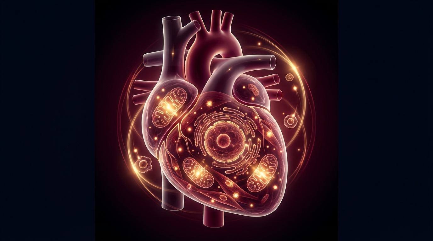
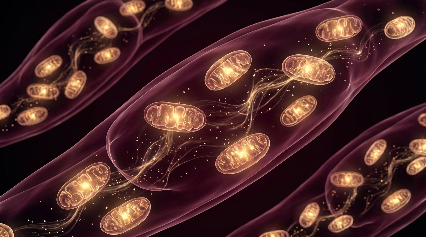
Every cell in the human body — with the exception of mature red blood cells — contains mitochondria. A single cell can house hundreds to thousands of them. They produce approximately 90% of the ATP that powers cellular function. When they work well, you feel it. When they do not, you feel that too.
Mitochondrial dysfunction is one of the 12 hallmarks of aging, but in my clinical experience, it is also one of the most practical targets for intervention. Patients with impaired mitochondrial function present with fatigue, cognitive decline, exercise intolerance, and metabolic disturbance — symptoms that are common, debilitating, and frequently dismissed as “normal aging.”
They are common. They should not be dismissed.
How Mitochondria Decline
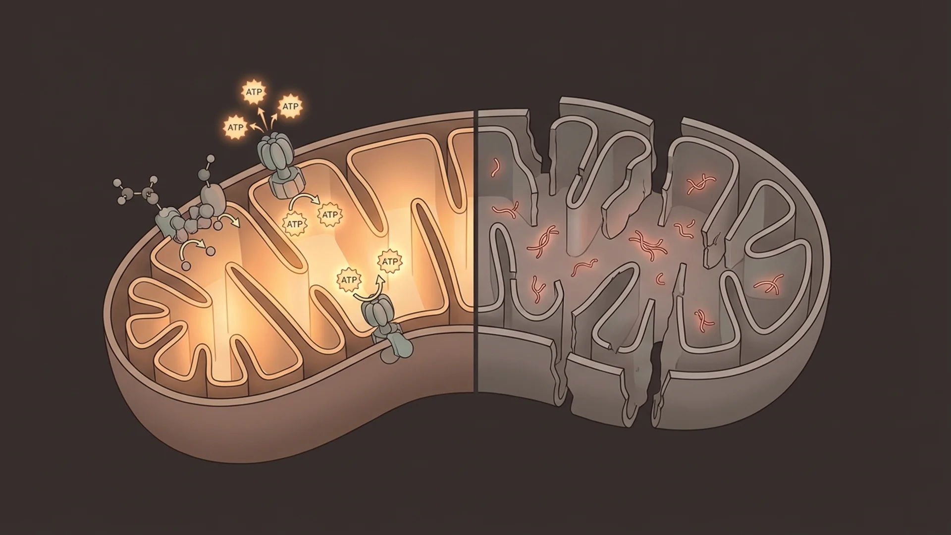
Mitochondria have their own DNA (mtDNA), inherited maternally, which encodes 13 essential components of the electron transport chain. This DNA is particularly vulnerable to damage because it lacks the robust repair mechanisms that protect nuclear DNA and sits in close proximity to the reactive oxygen species (ROS) that mitochondria produce as a byproduct of energy generation.
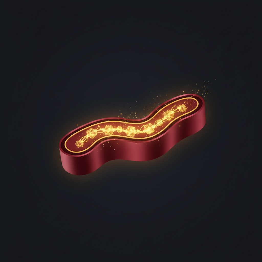
Over a lifetime, mtDNA mutations accumulate. The electron transport chain becomes less efficient. More ROS are produced. A vicious cycle establishes itself: damaged mitochondria produce more oxidative stress, which damages more mitochondria [1].
Additional drivers of mitochondrial decline include:
- NAD+ depletion — NAD+ is required for multiple steps in mitochondrial energy production. Its age-related decline directly impairs mitochondrial function.
- Alpha-ketoglutarate (AKG) depletion — this TCA cycle intermediate drops roughly tenfold between age 40 and 80, limiting the dioxygenase enzymes responsible for epigenetic maintenance and collagen synthesis. AKG supplementation may help restore this substrate deficit and works synergistically with NAD+ repletion.
- Reduced mitophagy — The selective autophagy of damaged mitochondria (mitophagy) declines with age, allowing dysfunctional organelles to persist — a process driven by PINK1/Parkin signalling that can be partially restored through fasting, exercise, urolithin A, and spermidine. Urolithin A, a gut-derived metabolite, is one of the few compounds shown in randomised trials to reactivate this PINK1/Parkin mitophagy pathway in humans.
- Environmental toxins — Heavy metals, pesticides, certain medications (notably statins and some antibiotics), and mold toxins can directly impair mitochondrial function.
- Chronic infection and inflammation — Persistent infections divert cellular resources and generate inflammatory mediators that impair mitochondrial efficiency.
Testing Mitochondrial Function
One of the challenges in mitochondrial medicine is that standard laboratory tests do not assess mitochondrial function directly. In my practice, I use a combination of approaches:
Organic acid testing provides indirect markers of mitochondrial function. Elevated citric acid cycle intermediates, abnormal ratios of specific organic acids, and markers of fatty acid oxidation can suggest mitochondrial impairment.
Lactate and pyruvate ratios offer a crude but clinically useful indicator. Elevated resting lactate or an abnormal lactate-to-pyruvate ratio can suggest impaired oxidative phosphorylation.
CoQ10 for mitochondrial support levels — Coenzyme Q10 is an essential electron carrier in the mitochondrial transport chain. Low levels correlate with impaired mitochondrial function and are common in patients on statin therapy. When prescribing CoQ10, the choice of form matters: Ubiquinol vs Ubiquinone: Which Form of CoQ10 Actually Works Better? covers the pharmacokinetic differences and when each form is appropriate.
MitoSwab and similar direct assessments — newer tests that assess mitochondrial complex function from buccal swabs are emerging, though validation data is still developing.
Functional assessment — sometimes the most informative approach is clinical. Exercise tolerance testing, perceived energy levels, and response to mitochondrial support therapies provide practical information that laboratory tests may miss.
Treatment: What Improves Mitochondrial Function
Foundational Interventions
Exercise is the most potent stimulus for mitochondrial biogenesis that we know of. Both aerobic exercise and resistance training trigger PGC-1alpha activation and the production of new, healthy mitochondria. High-intensity interval training appears particularly effective [2]. Zone 2 training is a particularly important foundation for mitochondrial health. For patients too fatigued to exercise, IHHT offers an alternative pathway to mitochondrial biogenesis without physical exertion.
Sleep is when much of the cellular repair and mitophagy occurs. Chronic sleep deprivation measurably impairs mitochondrial function.
Caloric restriction and time-restricted eating activate AMPK and sirtuins, both of which support mitochondrial quality control. Emerging research into mitochondria-encoded peptides — particularly MOTS-c, which activates AMPK via folate-cycle modulation — suggests that the mitochondrion itself produces longevity signals, and that these peptide levels decline measurably with age. For patients considering exogenous MOTS-c, our MOTS-c dosage and protocol guide covers injection schedules, cycling strategies, and clinical selection criteria in detail. Its sibling peptide humanin adds neuroprotective and insulin-sensitising dimensions to this same axis, with circulating levels falling roughly 40% between the third and eighth decades of life.
Targeted Supplementation
Coenzyme Q10 (CoQ10) — Ubiquinol, the reduced form, is essential for electron transport. Doses of 100-300 mg daily have shown benefit in controlled trials for heart failure (where the Q-SYMBIO data is compelling), statin-associated myopathy, and general fatigue [3]. In patients over 40, particularly those on statins, I consider CoQ10 assessment and supplementation routine.
NAD+ precursors or IV NAD+ — NAD⁺ decline is a primary upstream driver of mitochondrial dysfunction; restoring it via oral NMN or IV therapy addresses the substrate deficit directly. My NMN supplement dosing guide covers delivery forms, optimal dose ranges, and how to select the right approach based on clinical presentation.
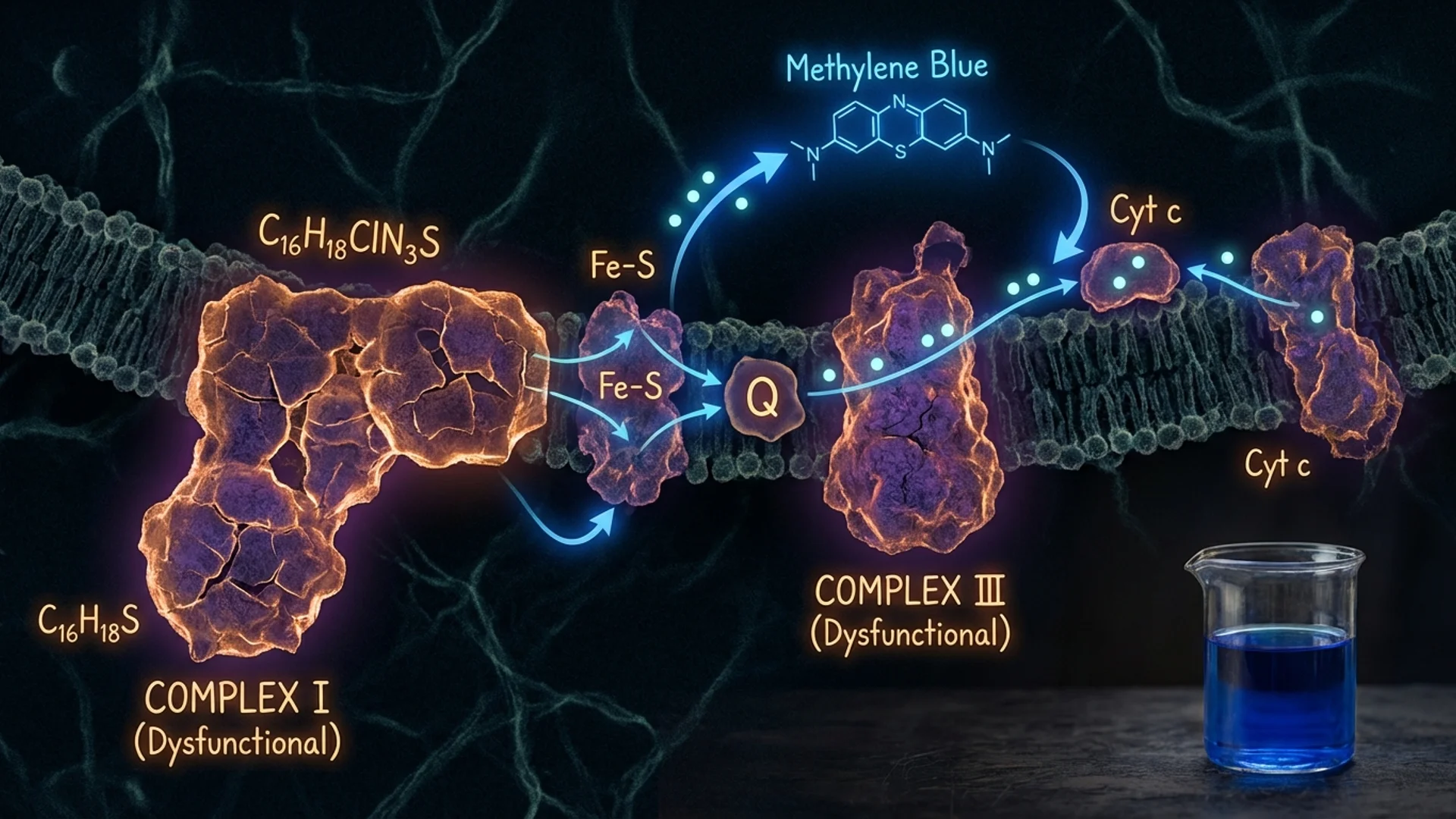
Shilajit (standardized for fulvic acid and dibenzo-α-pyrones) — A mineral-rich resin whose fulvic acid acts as an electron shuttle in Complex I and II, while its DBP fraction recycles ubiquinone back to active ubiquinol, effectively extending CoQ10’s functional lifespan. Particularly relevant in patients with post-viral fatigue, statin use, or age-related mitochondrial decline. Full clinical evidence review →
Alpha-lipoic acid (ALA) — A mitochondrial antioxidant that also supports glutathione recycling. Clinical trial data supports its use in diabetic neuropathy and general antioxidant support. Typical dose: 300-600 mg daily.
L-Ergothioneine — A histidine-derived amino acid found almost exclusively in mushrooms that your body maintains a dedicated transporter (OCTN1) to concentrate inside mitochondria-rich tissues. Unlike most antioxidants that are consumed on contact, ergothioneine cycles thousands of times before degradation, scavenging peroxynitrite and singlet oxygen directly at the source. Epidemiological data link lower plasma ergothioneine with faster cognitive decline and reduced longevity. Supplemental dose: 5–30 mg/day with food. Full clinical review →
Astaxanthin — A marine xanthophyll carotenoid with a unique molecular geometry that spans the full width of the mitochondrial membrane bilayer, providing antioxidant protection across both hydrophilic and hydrophobic zones simultaneously. Unlike alpha-lipoic acid or CoQ10, which operate in separate compartments, astaxanthin’s dual-surface protection is estimated to be 800× more potent than CoQ10 in singlet oxygen quenching assays. It also crosses the blood-brain barrier, making it especially relevant for patients with neurological and cognitive mitochondrial manifestations. Typical dose: 6–12 mg/day with a fat-containing meal. Full clinical review of astaxanthin for mitochondrial health.
GlyNAC (glycine + N-acetylcysteine) — Addresses glutathione depletion at its root by supplying both rate-limiting precursors simultaneously. A 24-week randomized controlled trial showed GlyNAC restored intracellular GSH to levels equivalent to young controls, normalized mitochondrial membrane potential, increased ATP production, and improved muscle strength, gait speed, and cognition in older adults. For patients with age-related mitochondrial decline, GlyNAC provides substrate-level support that indirect antioxidants cannot replicate. Clinical guide to GlyNAC dosing and evidence.
Acetyl-L-carnitine — Facilitates fatty acid transport into mitochondria for beta-oxidation. Particularly relevant for patients with fatigue and cognitive complaints. Doses of 500-2,000 mg daily are commonly used.
Sulforaphane — through NRF2/KEAP1 pathway activation — upregulates HO-1, NQO1, and glutathione synthesis enzymes, reducing the reactive oxygen species that directly damage mitochondrial DNA and drive the vicious cycle of mitochondrial decline. In patients with high oxidative load from chronic infection, toxin burden, or metabolic disease, sulforaphane works synergistically with CoQ10 and alpha-lipoic acid. See our full clinical review of sulforaphane and NRF2 for dosing strategies.
Molecular hydrogen (H₂ water) — A selective antioxidant that targets only the most cytotoxic free radicals — the hydroxyl radical and peroxynitrite — without dampening beneficial ROS signalling or blunting mitochondrial biogenesis. It also activates the Nrf2 pathway, similar to sulforaphane, boosting endogenous antioxidant enzyme expression. For patients with high oxidative burden or post-viral mitochondrial dysfunction, hydrogen-rich water is a practical, low-cost daily adjunct to IV NAD+ and CoQ10 protocols.
PQQ (pyrroloquinoline quinone) — Stimulates mitochondrial biogenesis through PGC-1alpha activation; human RCTs demonstrate improvements in cognitive performance and fatigue scores at 10–20 mg daily. Full clinical review and dosing guide. Typical dose: 10-20 mg daily.
TUDCA (tauroursodeoxycholic acid) — A water-soluble bile acid that stabilises the mitochondrial transition pore and attenuates all three arms of the unfolded protein response (UPR), preventing the shift from adaptive to pathological ER stress. It has demonstrated neuroprotection in ALS clinical trials and hepatoprotection in NASH RCTs. In patients with chronic infection, metabolic liver burden, or neurodegenerative concerns, TUDCA complements CoQ10 and NAD+ precursors at a structural level those molecules do not address. See the full TUDCA guide for clinical dosing.
B vitamins — Multiple B vitamins serve as cofactors in mitochondrial energy production. B1 (thiamine), B2 (riboflavin), B3 (niacin/niacinamide), and B5 (pantothenic acid) are all directly involved in the electron transport chain or citric acid cycle.
Magnesium — Required for ATP stabilization (ATP exists in cells as Mg-ATP). Deficiency is common and directly impairs energy production.
Clinical Interventions
IHHT — Intermittent Hypoxia-Hyperoxia Training stimulates mitochondrial turnover through controlled hypoxic stress. See my detailed article.
IV nutrient therapy — In patients with significant mitochondrial dysfunction, IV administration of key nutrients (NAD+, glutathione, B vitamins, magnesium, ALA) can bypass absorption limitations and achieve tissue levels not possible through oral supplementation alone.
Addressing root causes — Identifying and treating underlying contributors to mitochondrial dysfunction — chronic infections, heavy metal exposure, mold toxicity, medication effects — is essential. Supplementing mitochondria while the underlying insult continues is like bailing water without patching the hull.
Clinical Observations
In my practice, mitochondrial support is one of the most consistently rewarding areas of intervention. Patients with documented mitochondrial dysfunction who receive comprehensive treatment — combining lifestyle modification, targeted supplementation, and clinical therapies — frequently report meaningful improvements in energy, cognitive function, and exercise tolerance.
The improvements are not always dramatic or immediate. Mitochondrial repair takes time. I tell patients to expect gradual improvement over weeks to months, not overnight transformation.
What I have also observed is that mitochondrial dysfunction rarely exists in isolation. It typically coexists with hormonal imbalances, nutrient deficiencies, sleep disorders, or chronic infections. Addressing mitochondrial function as part of a comprehensive assessment produces better outcomes than treating it in isolation. When the clinical picture is dominated by acquired mitochondrial failure — particularly in patients with Lyme disease, post-COVID syndrome, or ME/CFS — a targeted diagnostic and treatment protocol becomes essential; our Mitochondrial Dysfunction: Root Causes, Testing, and Evidence-Based Treatment covers organic acids testing, intracellular nutrient panels, and layered recovery protocols in clinical detail.
The Bottom Line
Mitochondrial dysfunction is measurable, common, and treatable. It is not simply “getting older.” While we cannot fully reverse age-related mitochondrial decline, we can meaningfully support mitochondrial function through a combination of lifestyle interventions, targeted supplementation, and clinical therapies. For patients experiencing unexplained fatigue, cognitive decline, or exercise intolerance, mitochondrial assessment should be part of the evaluation.

References
- Sun N, Youle RJ, Bhargava P. The Mitochondrial Basis of Aging. Molecular Cell. 2016;61(5):654-666.
- Robinson MM, et al. Enhanced Protein Translation Underlies Improved Metabolic and Physical Adaptations to Different Exercise Training Modes in Young and Old Humans. Cell Metabolism. 2017;25(3):581-592.
- Mortensen SA, et al. The effect of coenzyme Q10 on morbidity and mortality in chronic heart failure: results from Q-SYMBIO. JACC Heart Failure. 2014;2(6):641-649.
This content is educational and does not constitute medical advice. Mitochondrial assessment and treatment should be supervised by a qualified physician.