
At a Glance
| Feature | Details |
|---|---|
| Class | Mitochondrial-derived peptide (MDP) |
| Length | 21 amino acids |
| Encoded by | Mitochondrial 16S rRNA gene |
| Primary receptors | gp130/CNTFR, FPRL1/FPR2 |
| Key actions | Neuroprotection, insulin sensitization, anti-apoptosis, anti-inflammation |
| Age-related trend | Declines ~40% between ages 20–80 |
| Clinical status | Experimental; no approved pharmaceutical form |
| Evidence level | Strong in vitro/animal; emerging human correlational data |
| Related MDPs | MOTS-C, SHLP1–6 |
In my clinical work with patients navigating chronic illness, complex metabolism, and accelerated aging, I encounter a recurring pattern: the mitochondria are almost always implicated. We discuss ATP production, reactive oxygen species, and the well-known supplement strategies — CoQ10, NAD precursors, PQQ. What fewer practitioners discuss is that mitochondria are not passive energy factories. They are active endocrine organs, secreting signaling molecules that coordinate survival responses throughout the body.
Humanin is the founding member of this class. Discovered in 2001 by Hashimoto and colleagues while screening for factors that rescue neurons from Alzheimer’s-associated cell death, humanin has since accumulated a body of evidence suggesting it is a master regulator of cellular resilience. Its decline with age may be one of the underappreciated contributors to neurodegeneration, metabolic disease, and reduced stress tolerance in older adults.
This article covers what humanin is, what the research shows about its physiological roles, how levels can be measured and influenced, and where it fits in an evidence-informed longevity protocol.
What Is Humanin and Where Does It Come From?
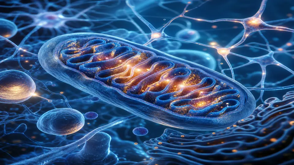
Humanin (HN) is a 21-amino-acid peptide encoded within the 16S ribosomal RNA region of mitochondrial DNA — making it one of a small family of open-reading-frame peptides transcribed from the mitochondrial genome rather than the nuclear genome. This is unusual; most proteins are nuclear-encoded. The fact that mitochondria retain this signaling capacity suggests deep evolutionary conservation.
The mature peptide is secreted from cells and circulates in plasma, acting in both autocrine (same cell) and endocrine (distant tissue) fashions. Its primary membrane receptors include the gp130/CNTFR complex — shared with CNTF, a well-characterized neurotrophic factor — and FPR2/FPRL1, a G-protein–coupled receptor also activated by anti-inflammatory lipoxins. These receptor targets immediately hint at humanin’s dual roles in neural protection and immune modulation.
The MDP Family Context
Humanin belongs to a growing family of mitochondria-derived peptides (MDPs) that also includes MOTS-C (which I have covered separately) and the six small humanin-like peptides (SHLP1–6). Each has distinct tissue distribution and functional emphasis:
- MOTS-C — metabolic regulation, AMPK activation, exercise mimicry
- SHLP2 and SHLP3 — mitochondrial function, cancer cell apoptosis
- Humanin — neuroprotection, insulin signaling, cardiovascular protection
Together, these peptides represent a previously unrecognized mitochondrial language by which our cells communicate stress and resilience signals. Age-related decline in their levels may be as important as the better-known hormonal declines in understanding why aging tissues lose function.
Humanin Levels Decline Predictably With Age
Cross-sectional human studies have measured circulating humanin by ELISA across age groups, and the findings are consistent: plasma humanin falls approximately 40% between the third and eighth decades of life. In the landmark 2016 study by Cobb and colleagues (PMID: 26962786), humanin levels in centenarians’ offspring were significantly higher than age-matched controls — suggesting that humanin preservation may be a feature of successful aging rather than a passive bystander.
Conditions associated with lower humanin levels include:
- Type 2 diabetes and insulin resistance
- Alzheimer’s disease and mild cognitive impairment
- Atherosclerosis and coronary artery disease
- Frailty and sarcopenia
- Chronic inflammatory states (elevated IL-6, TNF-α)
In my practice, when patients present with this cluster — metabolic dysfunction, cognitive complaints, cardiovascular risk, and chronic inflammation — I think of mitochondrial signaling capacity as a unifying thread.
Neuroprotective Mechanisms: The Original Discovery
The original 2001 paper by Hashimoto et al. (PMID: 11786514) identified humanin as a peptide that could rescue neuronal cell lines from death induced by multiple familial Alzheimer’s disease–associated genes (APP, presenilin-1, presenilin-2) and by amyloid-β itself. This finding was striking because no single factor had previously been shown to confer broad protection against such diverse neurotoxic insults.
Since then, humanin’s neuroprotective mechanisms have been characterized in considerable detail:
1. Inhibition of Bax-mediated mitochondrial apoptosis. Humanin binds directly to Bax, a pro-apoptotic BCL-2 family protein, preventing its translocation to the mitochondrial membrane and suppressing the cytochrome-c release cascade. This is a cell-autonomous survival mechanism.
2. Suppression of neuroinflammation. Via the FPR2 receptor and downstream NF-κB modulation, humanin reduces microglial activation and pro-inflammatory cytokine output. Given the central role of neuroinflammation in Alzheimer’s, Parkinson’s, and post-infectious brain fog, this pathway is clinically relevant.
3. STAT3-mediated trophic support. Signaling through gp130 activates STAT3, a transcription factor that upregulates neurotrophic genes including BDNF. This connects humanin to synaptic plasticity and neurogenesis.
4. Reduction of amyloid-β oligomer toxicity. Humanin can directly interact with Aβ aggregates, reducing their ability to form toxic oligomers. This raises the possibility that humanin decline contributes to amyloid pathology accumulation.
For patients presenting with post-COVID brain fog, Lyme-associated neurocognitive symptoms, or early cognitive decline, I consider the mitochondrial signaling axis — including humanin — as part of the biological picture alongside conventional workup.
Metabolic Effects: Insulin Sensitization and Glucose Homeostasis
A 2009 study by Muzumdar and colleagues (PMID: 19625522) demonstrated that centrally administered humanin dramatically improves peripheral insulin sensitivity in rodents — an effect mediated through hypothalamic STAT3 signaling rather than direct pancreatic action. Humanin-knockout mice develop insulin resistance, and restoration of humanin signaling rescues glucose metabolism.
In humans, cross-sectional analyses find that circulating humanin correlates inversely with fasting insulin, HOMA-IR (insulin resistance index), and visceral adiposity. Patients with type 2 diabetes have consistently lower humanin levels than matched controls.
The proposed mechanisms include:
- Central sensitization of hypothalamic insulin circuits
- Suppression of hepatic glucose output via STAT3 in hepatocytes
- Anti-inflammatory effects that reduce cytokine-driven insulin resistance
- Mitochondrial protection in pancreatic beta cells, preserving insulin secretory capacity
This metabolic profile places humanin alongside MOTS-C and other MDPs as part of the reason exercise and caloric restriction improve insulin sensitivity beyond their known mechanical effects — both interventions upregulate MDP expression.
Cardiovascular Protection
Cardiomyocytes are among the most mitochondria-dense cells in the body, and humanin exerts direct protective effects on heart tissue. Kim and colleagues (PMID: 29355857) showed that humanin inhibits atherosclerotic plaque progression in ApoE-knockout mice by reducing foam cell formation and suppressing vascular inflammation.
Key cardiovascular mechanisms:
- Anti-apoptotic effect in ischemia-reperfusion injury: Humanin pre-treatment reduces cardiomyocyte death when blood flow is experimentally restored after occlusion — the “reperfusion injury” paradigm relevant to post-MI tissue loss
- Endothelial protection: Humanin suppresses oxidized-LDL–induced endothelial cell death and adhesion molecule expression
- Platelet-activating factor antagonism: Via FPR2 receptor signaling, humanin partially antagonizes platelet-activating factor, a pro-thrombotic and pro-inflammatory mediator
In the centenarian offspring study cited above, higher humanin levels were associated with better lipid profiles, lower CRP, and lower cardiovascular event rates — consistent with these mechanistic findings.
How to Support Humanin Levels: Lifestyle and Therapeutic Strategies
No pharmaceutical humanin preparation is currently approved or available through regulated compounding in most jurisdictions. However, several strategies are supported by evidence to influence humanin expression or replicate aspects of its signaling:
Lifestyle Approaches
| Strategy | Mechanism | Evidence |
|---|---|---|
| High-intensity interval training | Mitochondrial biogenesis, upregulates MDP expression | Strong |
| Caloric restriction / intermittent fasting | Mitochondrial stress hormesis, SIRT1 activation | Moderate |
| Heat exposure (sauna) | Mitochondrial unfolded protein response | Preliminary |
| Cold plunge | Mitochondrial ROS signaling, PGC-1α activation | Preliminary |
Supplement Stack Supporting MDP Signaling
- NAD+ precursors (NMN, NR): Sirtuins regulate mitochondrial biogenesis and MDP expression; NAD+ repletion supports this axis
- CoQ10/Ubiquinol: Maintains electron transport chain function, reducing mitochondrial stress that depletes MDPs
- Alpha-ketoglutarate: Supports mitochondrial TCA cycle function and has shown longevity effects in animal models
- Spermidine: Induces mitophagy, clearing dysfunctional mitochondria and stimulating biogenesis
- SS-31 (elamipretide): A mitochondria-targeted tetrapeptide that works at the structural level — binding cardiolipin on the inner mitochondrial membrane to restore cristae architecture and reduce electron leak. Where humanin acts through receptor signalling, SS-31 repairs the membrane platform that respiratory supercomplexes depend on — the two approaches are complementary.
Investigational Approaches
Synthetic humanin analogues — particularly HNG (humanin with glycine substitution at position 14) — show 1,000-fold greater potency than native humanin in preclinical models. HNG is used in research protocols but is not yet available clinically. Several biotech companies are developing humanin-based therapeutics for Alzheimer’s prevention and metabolic disease, and early-phase trials are underway.
Practitioners working with exosome therapies should also note that mesenchymal stem cell–derived exosomes contain MDPs including humanin, which may partly explain their observed neuroprotective and metabolic effects in some clinical series.
Measuring Humanin Clinically
Plasma humanin can be quantified via ELISA (enzyme-linked immunosorbent assay) at specialty reference laboratories, though it is not yet a standard panel in most health systems. Reference ranges are not fully established, but published cohort data suggest:
- Young adults (20–40 years): ~1.5–3.0 ng/mL plasma
- Middle age (40–60 years): ~1.0–2.0 ng/mL
- Older adults (60–80 years): ~0.8–1.5 ng/mL
- Centenarian offspring: modestly but significantly higher than age-matched controls
In my practice, I include humanin alongside MOTS-C and other mitochondrial function markers — such as cardiolipin antibodies, organic acids panel (for mitochondrial metabolite profiling), and lactate-to-pyruvate ratio — when building a comprehensive mitochondrial health picture for patients with complex chronic disease or accelerated aging presentation.
Clinical Perspective: Where Humanin Fits
Humanin is not a standalone therapy today, but it represents a paradigm shift in how we understand aging biology. The finding that our own mitochondria encode survival signals — and that these signals predictably decline with age — adds a new dimension to longevity medicine beyond hormones, lifestyle, and conventional supplements.
For my patients, the practical takeaway is this: protect your mitochondria and they protect you. The strategies that keep mitochondria healthy — adequate NAD+ substrate, exercise-induced hormesis, strategic caloric restriction, targeted supplementation, and reduced toxic burden — are the same strategies that support humanin and the broader MDP axis.
As the synthetic analogue pipeline matures, I expect humanin-based interventions to become a meaningful tool in neuroprotection protocols, particularly for patients with early cognitive changes or high Alzheimer’s risk. I am watching the investigational HNG literature closely and will update this guidance when clinical-grade preparations become available.
Related Articles
- MOTS-C Peptide: Metabolic Regulation and Exercise Mimicry
- NAD+ Supplement Guide: NMN, NR, and IV Options Compared
- Mitochondrial Health: Why Your Power Plants Determine Your Longevity
- Peptide Therapy for Women: Hormones, Healing, and Longevity
- Longevity Supplement Stack: A Physician’s Evidence-Based Protocol
References
- Hashimoto Y, et al. A rescue factor abolishing neuronal cell death by a wide spectrum of familial Alzheimer’s disease genes and Abeta. Proc Natl Acad Sci USA. 2001;98(11):6336–6341. PMID: 11786514
- Cobb LJ, et al. Naturally occurring mitochondrial-derived peptides are age-dependent regulators of apoptosis, insulin sensitivity, and inflammatory markers. Aging (Albany NY). 2016;8(4):796–809. PMID: 26962786
- Muzumdar RH, et al. Humanin: a novel central regulator of peripheral insulin action. PLoS ONE. 2009;4(7):e6334. PMID: 19625522
- Kim SJ, et al. Mitochondria-derived peptides as novel regulators of cardiometabolic function. Annu Rev Physiol. 2018;80:281–307. PMID: 29355857
- Lee C, et al. The mitochondrial-derived peptide MOTS-c promotes metabolic homeostasis and reduces obesity and insulin resistance. Cell Metab. 2015;21(3):443–454. PMID: 25738459
- Guo B, et al. Humanin peptide suppresses apoptosis by interfering with Bax activation. Nature. 2003;423(6938):456–461. PMID: 12721631
- Cohen P, Bhargava G, Bhargava A. New roles for humanin in the mitochondrial unfolded protein response and cytoprotection. Ageing Res Rev. 2020;64:101197. PMID: 33011326
For a detailed breakdown of dosing protocols, administration routes, cycling schedules, and stacking combinations, see the companion Humanin Peptide Dosage Protocol guide.