At a Glance
| Property | Value |
|---|---|
| Evidence Level | Moderate |
| Primary Use | Understanding the neurobiological basis of cognitive dysfunction in tick-borne disease |
| Key Mechanism | Microglial activation producing neuroinflammatory mediators that disrupt synaptic function, neurotransmitter metabolism, and cerebral blood flow |
Brain Fog Is Not in Your Head — It Is in Your Brain
If you have Lyme disease and brain fog, you have probably been told some version of “your scans look normal” or “this is probably stress-related.” Standard MRI and CT imaging typically appear unremarkable in Lyme patients with cognitive dysfunction, leading to the dismissal of a symptom that patients describe as one of the most disabling features of their illness.
Here is what I tell my patients: standard brain imaging is not designed to detect neuroinflammation. It shows structure, not inflammation. The fact that your MRI is normal does not mean your brain is normal — it means the tool being used is not sensitive enough to detect what is happening.
The research that changed this conversation came from Johns Hopkins.
The Johns Hopkins PET Scan Study
Researchers at the Johns Hopkins Lyme Disease Research Center used [11C]DPA-713 PET (positron emission tomography) — a specialized imaging technique that detects translocator protein (TSPO) expression on activated microglial cells — to image the brains of patients with post-treatment Lyme disease syndrome (PTLDS) [1].
What They Found
Compared to healthy controls, PTLDS patients showed significantly elevated TSPO signal in multiple brain regions, indicating widespread microglial activation and neuroinflammation. The key findings:
- Widespread glial activation across multiple brain regions, not limited to a single area
- Correlation with symptoms — the severity of neuroinflammation correlated with the severity of self-reported cognitive symptoms
- Persistence after treatment — the neuroinflammation was present in patients who had completed standard antibiotic therapy, suggesting that the inflammatory process continues beyond active infection
This was the first study to provide direct neuroimaging evidence that brain fog in Lyme disease has a measurable biological correlate — neuroinflammation is real, visible, and quantifiable.
Why This Matters
The significance of this research cannot be overstated for patients who have been told their cognitive symptoms are psychological:
- Validation: Brain fog in Lyme disease is not imagined, exaggerated, or stress-related. It has an identifiable neurobiological mechanism visible on appropriate imaging.
- Treatment direction: If brain fog is caused by neuroinflammation, treatment should target neuroinflammation — not just the infection.
- Persistence explanation: The continued presence of neuroinflammation after antibiotic therapy explains why brain fog often persists even when other Lyme symptoms improve.
The Mechanism: How Neuroinflammation Creates Brain Fog
Microglial Activation
Microglia are the resident immune cells of the central nervous system. In their resting state, they perform surveillance and maintenance functions. When activated by infection, injury, or biotoxins, they transform into a pro-inflammatory phenotype — producing TNF-alpha, IL-1beta, IL-6, reactive oxygen species, and nitric oxide [2].
In Lyme disease, microglia can be activated by:
- Direct Borrelia invasion of the CNS (neuroborreliosis)
- Borrelia outer surface proteins crossing the blood-brain barrier
- Systemic inflammatory mediators reaching the brain through a compromised blood-brain barrier
- Reactivated viruses with CNS tropism (particularly HHV-6)
Neurotransmitter Disruption
Activated microglia alter the kynurenine pathway — the primary metabolic route for tryptophan, the amino acid precursor to serotonin. Instead of producing serotonin, inflamed brain tissue shunts tryptophan toward quinolinic acid — a neurotoxic metabolite that:
- Overstimulates NMDA receptors (excitotoxicity)
- Depletes serotonin (contributing to mood dysfunction)
- Generates free radicals (oxidative neuronal damage)
- Impairs synaptic plasticity (reducing learning and memory)
This shift explains why Lyme patients with brain fog often also experience mood changes, sleep disruption, and anxiety — they share a common neuroinflammatory mechanism.
Impaired Cerebral Blood Flow
Neuroinflammation impairs cerebral autoregulation — the brain’s ability to maintain consistent blood flow across varying blood pressures. This is compounded by the autonomic dysfunction common in chronic Lyme patients, which further impairs cerebral perfusion.
SPECT (Single Photon Emission Computed Tomography) imaging in Lyme patients has consistently shown regional hypoperfusion — areas of reduced blood flow — in temporal and frontal lobes. These are the regions responsible for memory, executive function, and processing speed — the exact cognitive domains that brain fog affects.
White Matter Changes
While standard MRI may appear normal, advanced MRI techniques (diffusion tensor imaging, DTI) have detected white matter abnormalities in PTLDS patients — suggesting disruption of the myelinated pathways connecting different brain regions. This is consistent with both direct inflammatory damage and HHV-6 infection of oligodendrocytes (the cells that produce myelin).

The Evidence
What We Know (Human Data)
The Johns Hopkins PET scan study (Coughlin et al.) represents the strongest direct evidence for neuroinflammation in PTLDS. Additional supporting evidence includes:
- SPECT imaging studies documenting cerebral hypoperfusion in Lyme patients (Logigian et al., Fallon et al.)
- Neuropsychological testing consistently demonstrating processing speed, working memory, and executive function deficits in PTLDS patients [3]
- CSF biomarker studies showing elevated inflammatory markers (CXCL13, IL-6, neopterin) in neuroborreliosis patients
- Cognitive improvement with anti-inflammatory treatment in multiple case series
The evidence is moderate — consistent across multiple modalities but limited by small sample sizes and the absence of large multicenter trials.
What I See in Practice
In our clinical experience, brain fog is the most common complaint among our chronic Lyme patients — present in over 80% of cases. And it is consistently the symptom that persists longest, often remaining after joint pain, fatigue, and other systemic symptoms have improved.
What I observe in practice is that brain fog responds to a different treatment approach than the systemic infection. Anti-Borrelia therapy reduces the upstream trigger but often does not fully resolve the neuroinflammation. The patients who experience the most complete cognitive recovery are those who receive targeted neuroinflammation treatment in addition to antimicrobials.
I also observe that brain fog severity correlates strongly with two factors:
- Duration of illness — patients sick for years have more entrenched neuroinflammation than those treated within months
- Viral co-reactivation — patients with reactivated EBV and/or HHV-6 have significantly worse brain fog than those without viral reactivation
Practical Application
Diagnosing Neuroinflammation
Standard neurological workup (MRI, EEG, basic neuropsych) will typically be normal or mildly abnormal. More sensitive approaches:
- Neuropsychological testing — quantifies processing speed, working memory, and executive function deficits
- SPECT imaging — detects cerebral hypoperfusion patterns
- TSPO PET (research setting only, not widely available) — direct neuroinflammation imaging
- CSF analysis — if lumbar puncture is clinically indicated: CXCL13, white cell count, protein, Borrelia antibody index
Treatment Strategy for Neuroinflammation-Mediated Brain Fog
1. Treat the upstream trigger
- Continue anti-Borrelia therapy
- Address viral co-reactivation (EBV, HHV-6)
- Address mold/CIRS if present
- Optimize the vagus nerve pathway to modulate neuroinflammation
2. Direct anti-neuroinflammatory therapy
- Low-dose naltrexone (LDN) 1.5-4.5mg nightly — evidence for microglial modulation in neuroinflammatory conditions
- Curcumin (bioavailable form) — crosses BBB, inhibits NF-kB, reduces microglial activation
- Omega-3 fatty acids (high-dose EPA) — specialized pro-resolving mediators that actively resolve neuroinflammation
- N-acetylcysteine (NAC) — reduces oxidative stress and quinolinic acid-mediated excitotoxicity
- Palmitoylethanolamide (PEA) — endogenous anti-neuroinflammatory lipid; 600mg twice daily
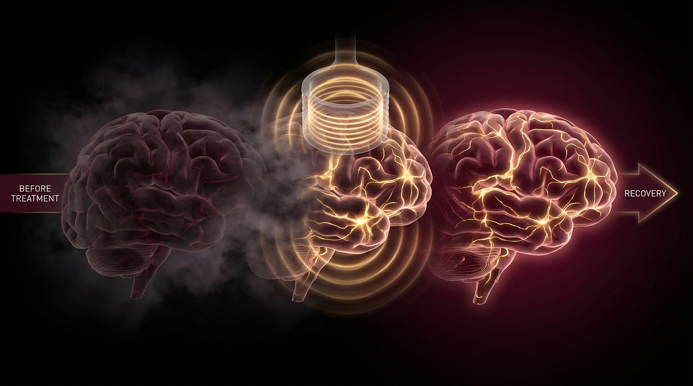
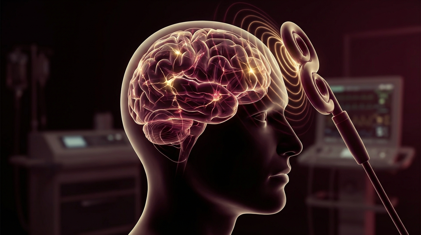
3. Neuromodulation approaches
- Transcranial Pulse Stimulation (TPS) — the approach we are most confident in for neurological recovery
- Vagus nerve stimulation — activates the cholinergic anti-inflammatory pathway
- PEMF therapy — evidence for reducing neuroinflammation and promoting neuronal recovery
- Neurofeedback — retrains brainwave patterns disrupted by chronic neuroinflammation
4. Cerebrovascular support
- Ginkgo biloba — evidence for improving cerebral perfusion (EGb 761 standardized extract)
- Vinpocetine — enhances cerebral blood flow and oxygen utilization
- Exercise — the most evidence-supported intervention for improving cerebral perfusion (start with vagus nerve exercises if autonomic dysfunction prevents traditional exercise)
5. Brain fog recovery timeline — set realistic expectations. Neuroinflammation resolves over months, not weeks.

Safety and Considerations
- Low-dose naltrexone is generally well-tolerated but should not be used with opioid medications. Start at 1.5mg and titrate to 4.5mg over 2-4 weeks.
- Not all brain fog is neuroinflammation. Sleep deprivation, medication side effects, hormonal dysfunction, nutritional deficiencies, and primary psychiatric conditions should be evaluated.
- Neuropsychological testing provides objective measurement of cognitive deficits — valuable both for diagnosis and for tracking treatment response.
- Brain fog recovery takes time. Patients should expect gradual improvement over 3-12 months, not rapid resolution. Neuroinflammation that has been present for years does not resolve in weeks.
- The PET scan findings from Johns Hopkins are promising but based on a small cohort. Larger replication studies are needed to confirm and expand these findings.
The Bottom Line
Brain fog in Lyme disease is neuroinflammation — measurable, visible on appropriate imaging, and mechanistically understood. The Johns Hopkins PET scan research proved that microglial activation persists in PTLDS patients, providing biological validation for a symptom that patients have been told is “in their head.” Treatment must go beyond antimicrobials: targeted anti-neuroinflammatory therapy, neuromodulation, cerebrovascular support, and addressing viral co-reactivation are essential for cognitive recovery. In my clinical experience, patients who receive comprehensive neuroinflammation-directed treatment achieve significantly better cognitive outcomes than those treated with antimicrobials alone.
References
- Coughlin JM, Yang T, Rebman AW, et al. Imaging glial activation in patients with post-treatment Lyme disease symptoms: a pilot study using [11C]DPA-713 PET. J Neuroinflammation. 2018;15(1):346. PMID: 30526632
- Salter MW, Stevens B. Microglia emerge as central players in brain disease. Nat Med. 2017;23(9):1018-1027. PMID: 28886007
- Rebman AW, Bechtold KT, Yang T, et al. The clinical, symptom, and quality-of-life characterization of a well-defined group of patients with posttreatment Lyme disease syndrome. Front Med. 2017;4:224. PMID: 29312942
- Fallon BA, Keilp JG, Corbera KM, et al. A randomized, placebo-controlled trial of repeated IV antibiotic therapy for Lyme encephalopathy. Neurology. 2008;70(13):992-1003. PMID: 17928580