At a Glance
| Property | Value |
|---|---|
| Evidence Level | Moderate |
| Primary Use | Diagnosis and management of reactivated Epstein-Barr virus |
| Key Mechanism | Immune suppression allows latent EBV to reactivate from B-lymphocyte reservoir, driving fatigue through mitochondrial dysfunction and immune dysregulation |
The Virus That Never Left
You had mono in college — or maybe you never noticed the primary infection at all. Either way, Epstein-Barr virus has been living quietly in your B-lymphocytes since that initial exposure, held in check by your immune system. For most of your life, this arrangement worked perfectly.
Then something changed. Maybe you developed Lyme disease. Maybe you moved into a water-damaged building. Maybe you went through a period of extreme stress, surgery, or another infection. Whatever it was, your immune surveillance dropped — and EBV woke up.
Here is what the evidence shows about why this happens, how to identify it, and what to do about it.
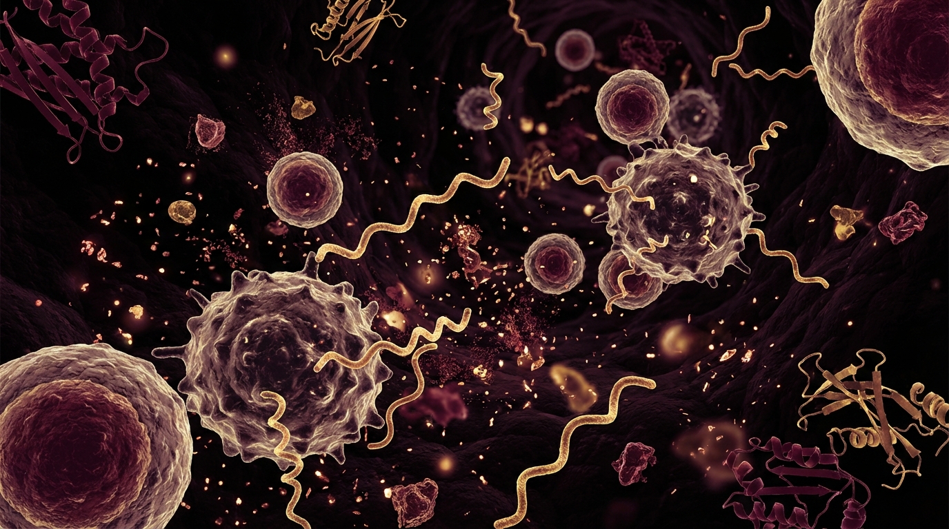
The Biology of EBV Latency and Reactivation
Epstein-Barr virus (HHV-4) infects over 90% of the global adult population. After primary infection (which may manifest as infectious mononucleosis or be entirely subclinical), the virus establishes lifelong latency in memory B-lymphocytes [1].
During latency, EBV expresses only a minimal set of genes — enough to maintain its presence in B-cells but not enough to trigger immune detection. This is an evolutionarily elegant strategy: the virus hides in the very cells that should be detecting it.
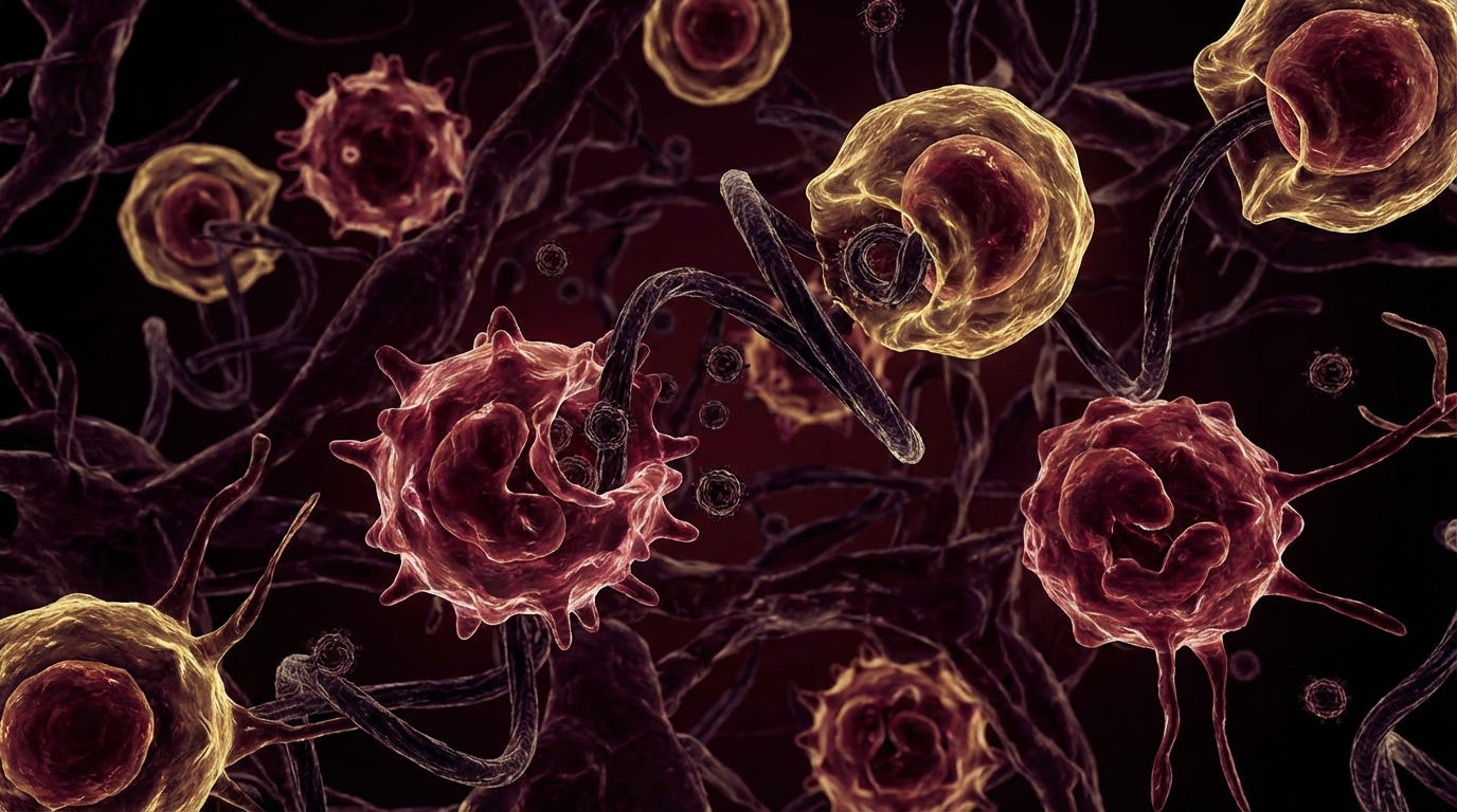
What Triggers Reactivation
EBV reactivation occurs when the T-cell and NK-cell surveillance that keeps the virus latent is compromised. Documented triggers include:
- Chronic infections: Lyme disease, Bartonella, other tick-borne infections — these suppress NK cell and CD8+ T-cell function
- Mold/CIRS: Biotoxin illness depletes MSH and impairs immune regulation
- Chronic stress: Cortisol suppresses cellular immunity, particularly NK cell cytotoxicity
- Immunosuppressive medications: Steroids, biologics, transplant drugs
- Surgery or physical trauma: Transient immune suppression
- Other viral infections: COVID-19 has been documented to trigger EBV reactivation
- Aging: Immunosenescence reduces T-cell surveillance efficiency
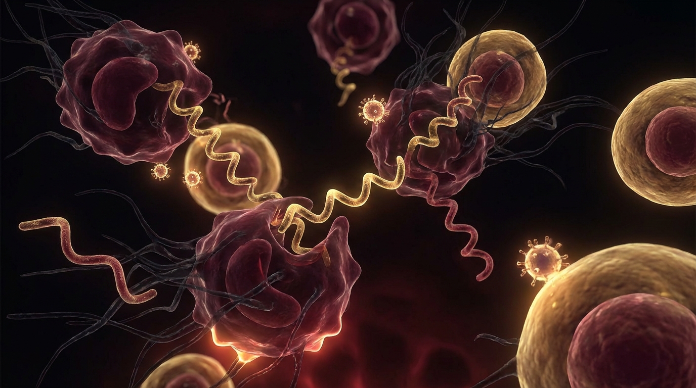
The Reactivation Cascade
When EBV reactivates, it transitions from latent to lytic (active replication) phase:
- Viral gene expression shifts from latent (EBNA, LMP) to lytic (early antigen, viral capsid antigen)
- New virions are produced, infecting additional B-cells
- Infected cells produce viral IL-10 (vIL-10) — a viral mimic of human IL-10 that suppresses the immune response
- Activated B-cells produce pro-inflammatory cytokines, contributing to fatigue and malaise
- Mitochondrial function is disrupted through direct viral effects and cytokine-mediated damage
- The resulting immune dysregulation can destabilize other latent viruses (HHV-6, CMV, VZV)
Symptoms of Reactivated EBV
Reactivated EBV does not look like acute mono in most cases. The presentation is typically more subtle and chronic:
Primary symptoms:
- Profound, disproportionate fatigue (the hallmark)
- Sore throat (often mild but persistent)
- Tender cervical lymph nodes
- Low-grade fever or temperature dysregulation
- Brain fog and cognitive dysfunction
- Muscle and joint pain
Associated features:
- Night sweats
- Headaches
- Sleep disruption (unrefreshing sleep)
- Liver enzyme elevation (mild)
- Splenomegaly (less common than in primary infection)
What distinguishes reactivated EBV from primary mononucleosis is the predominance of fatigue over acute symptoms. Patients with reactivation rarely have the dramatic pharyngitis and lymphadenopathy of acute mono. Instead, they have a grinding, relentless exhaustion that does not respond to rest.

Diagnosis: Getting the Serology Right
This is where many physicians make errors. Standard EBV testing is designed to distinguish past infection from acute primary infection — not to detect reactivation. Understanding the serology is critical.
| Marker | Past Infection (Dormant) | Acute Primary Infection | Reactivation |
|---|---|---|---|
| VCA IgM | Negative | Positive | Positive or borderline |
| VCA IgG | Positive | Positive | Positive (often high) |
| EA (Early Antigen) IgG | Negative | Positive (early) | Positive |
| EBNA IgG | Positive | Negative (develops late) | Positive |
The key markers for reactivation:
- EA (Early Antigen) IgG elevation — this is the most important marker. EA IgG is produced during active viral replication and should be low or negative in latent infection. Elevated EA IgG in the setting of positive EBNA IgG suggests reactivation from prior latent infection [1].
- VCA IgM re-emergence — VCA IgM normally appears during primary infection and then declines. Its reappearance (or persistent positivity) suggests reactivation.
- Quantitative EBV PCR — detects viral DNA in blood, confirming active viral replication. This is the most definitive test for reactivation.
The common error: A physician orders “EBV panel,” sees positive VCA IgG and positive EBNA IgG, and tells the patient “you had EBV in the past, it is not active.” This interpretation misses the EA IgG result entirely and does not consider the possibility of reactivation. If your EA IgG is elevated and you have compatible symptoms, your EBV is likely reactivated regardless of what the VCA IgG and EBNA show.
The Evidence
What We Know (Human Data)
EBV reactivation has been documented extensively in immunocompromised populations — transplant recipients, HIV patients, and cancer patients receiving chemotherapy. In these populations, reactivation can be severe (post-transplant lymphoproliferative disorder, EBV-associated lymphomas) [2].
In the context of ME/CFS, a growing body of research supports EBV reactivation as a contributing factor. A 2019 study in Frontiers in Immunology demonstrated that ME/CFS patients had significantly higher EBV reactivation markers compared to healthy controls, and that this reactivation correlated with NK cell dysfunction [3].
The COVID-19 pandemic has added substantial evidence: multiple studies documented EBV reactivation during and after SARS-CoV-2 infection, with reactivation associated with Long COVID symptom severity. Gold et al. (2021) found that 66.7% of Long COVID patients had evidence of EBV reactivation compared to 10% of controls.
What I See in Practice
In our clinical experience, EBV reactivation is present in the majority of our chronic Lyme patients — I would estimate 60-70% show laboratory evidence of reactivation. This is not surprising: Borrelia and its co-infections suppress the very immune pathways (NK cells, CD8+ T-cells) responsible for maintaining EBV latency.
What I observe in practice is that treating only the tick-borne infections without addressing the reactivated EBV often produces incomplete recovery. The patient improves with anti-Borrelia therapy but continues to have disproportionate fatigue and cognitive dysfunction. When we add antiviral therapy for EBV, an additional layer of improvement typically follows.
I also observe that the patients who struggle most with EBV reactivation are those with concurrent mold/CIRS. The biotoxin burden further suppresses immune surveillance, creating a three-way problem: mold → immune suppression → EBV reactivation → further immune suppression → Lyme treatment failure.
Practical Application
Treatment Strategy
1. Address the underlying immune stressor (always first)
- Treat the primary infection (Lyme, Bartonella, Babesia)
- Address mold exposure and CIRS if present
- Optimize sleep, stress management, nutrition
- This step is essential — treating EBV without removing the trigger for reactivation produces only temporary improvement
2. Antiviral therapy
- Valacyclovir 1g three times daily — the most commonly used antiviral for EBV reactivation in clinical practice
- Duration: 3-6 months minimum for chronic reactivation
- Note: Valacyclovir primarily inhibits viral DNA polymerase during lytic replication; it does not eliminate latent virus
- Some clinicians use famciclovir as an alternative
3. Immune support
- Thymosin alpha-1 — enhances T-cell and NK cell function; FDA-approved in 35+ countries for immune modulation
- Vitamin D optimization — target 50-80 ng/mL; vitamin D regulates antimicrobial peptide production and NK cell activity
- Zinc — 30-50mg daily; essential for T-cell maturation and function
- Medicinal mushrooms — AHCC, turkey tail (Trametes versicolor), reishi — evidence supporting NK cell enhancement
- Selenium — 200mcg daily; supports antiviral immune response
4. Mitochondrial support
- EBV reactivation impairs mitochondrial function through direct viral effects and cytokine-mediated damage
- CoQ10 200-400mg daily (ubiquinol form)
- NAD+ support — NMN or NR supplementation; IV NAD+ in clinical settings
- B vitamins — methylated forms (methylfolate, methylcobalamin)
5. Monitor and reassess
- Repeat EBV serology (EA IgG, VCA IgM) every 3 months
- Quantitative EBV PCR to track viral load
- NK cell function testing to assess immune recovery
- Symptom tracking with standardized questionnaire

Safety and Considerations
- Valacyclovir is generally well-tolerated but can cause headache, nausea, and renal impairment at higher doses. Adequate hydration and periodic renal function monitoring are recommended.
- EBV reactivation can drive or worsen autoimmune conditions (multiple sclerosis, lupus, rheumatoid arthritis). Persistent reactivation warrants monitoring for autoimmune markers.
- Not all elevated EA IgG indicates clinically significant reactivation. Interpretation requires clinical correlation with symptoms and additional markers.
- Immunosuppressive medications (including biologics for autoimmune disease) can both trigger and mask EBV reactivation. Close monitoring is essential in these patients.
- Herxheimer-like reactions can occur when initiating antiviral therapy, though less commonly than with antibacterial treatment.
The Bottom Line
EBV reactivation is one of the most common and most overlooked contributors to chronic fatigue in patients with tick-borne diseases, mold illness, and post-COVID syndrome. The virus that 90% of us carry dormantly can become a significant disease driver when immune surveillance fails. Diagnosis requires looking beyond the standard EBV panel — EA IgG and EBV PCR are the critical markers. Treatment requires a two-pronged approach: antiviral therapy for the virus itself, and restoration of the immune surveillance that should be keeping it dormant. In my clinical experience, patients who address both the EBV and its underlying trigger have markedly better outcomes than those who treat only one.
References
- Houen G, Trier NH. Epstein-Barr virus and systemic autoimmune diseases. Front Immunol. 2021;11:587380. PMID: 33488588
- Kerr JR. Epstein-Barr virus (EBV) reactivation and therapeutic inhibitors. J Clin Pathol. 2019;72(10):651-658. PMID: 31315893
- Loebel M, Strohschein K, Giannini C, et al. Deficient EBV-specific B- and T-cell response in patients with chronic fatigue syndrome. PLoS One. 2014;9(1):e85387. PMID: 24416400
- Gold JE, Okyay RA, Licht WE, Hurley DJ. Investigation of Long COVID prevalence and its relationship to Epstein-Barr virus reactivation. Pathogens. 2021;10(6):763. PMID: 34204243