At a Glance
| Feature | Detail |
|---|---|
| Primary mechanism | Extracorporeal removal of autoantibodies, immune complexes, and inflammatory mediators |
| Session duration | 2–3 hours per session |
| Typical course | 5–10 sessions over 1–2 weeks |
| Best-responding conditions | Myasthenia gravis, rheumatoid arthritis, lupus nephritis, MCAS, CIDP |
| Risk profile | Low in monitored clinical setting; mild fatigue and transient hypotension most common |
| Evidence tier | ASFA Category I–II for established indications; extensive clinical series for complex autoimmune overlap |
When a patient’s immune system turns against itself, the standard medical response is to suppress it systemically — with corticosteroids, disease-modifying antirheumatic drugs (DMARDs), or biologics. This approach manages symptoms effectively for many people. For those who fail multiple lines of therapy, or who cannot carry the side-effect burden of long-term immunosuppression, a different logic applies: rather than suppressing the immune system, remove the offending molecules from circulation entirely.
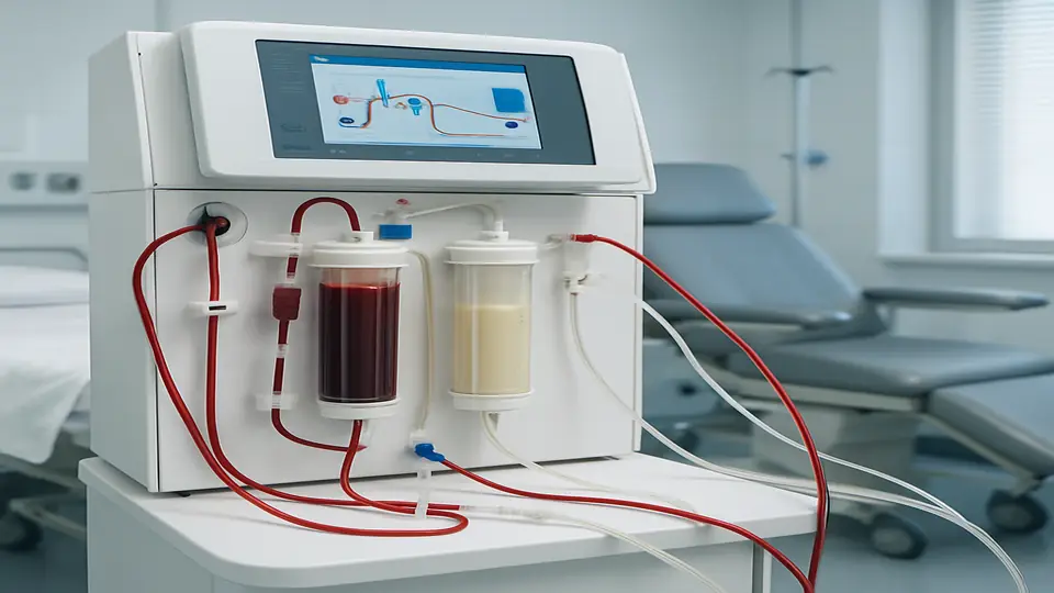
Therapeutic apheresis does exactly that. By routing blood through an extracorporeal circuit and passing it through specialized filters or adsorption columns, clinicians can selectively reduce the circulating load of autoantibodies, immune complexes, and pro-inflammatory cytokines — in a matter of hours. For patients whose autoimmune disease has escaped pharmacological control, this can be genuinely transformative.
How Apheresis Works: The Extracorporeal Approach
Apheresis (from the Greek aphairein — to take away) is a family of blood purification techniques in which blood is drawn from the patient, processed through an external device, and returned to the body. The relevant modalities for autoimmune disease include several distinct approaches, each with different targets and selectivity profiles.
Therapeutic plasma exchange (TPE) separates the entire plasma fraction and discards it, replacing it with albumin or fresh frozen plasma. This removes large-molecular-weight proteins non-selectively, including immunoglobulins, complement proteins, and immune complexes. TPE is well-established for acute indications but depletes beneficial proteins alongside pathological ones.
Immunoadsorption (IAS) is the more selective approach. Plasma is passed through a column containing ligands specific to IgG antibodies — tryptophan, protein A, or phenylalanine-based matrices. This removes immunoglobulins, including pathogenic autoantibodies, with substantially less depletion of albumin and clotting factors compared to plasma exchange. The ability to process larger plasma volumes per session without needing replacement fluid makes immunoadsorption particularly attractive for outpatient protocols.
HELP apheresis and Inuspheresis extend this further. These systems target not only immunoglobulins but also lipoprotein-bound inflammatory mediators, fibrinogen, and cytokine-carrying particles, with documented effects on oxidative stress, microcirculatory function, and vascular inflammation. For patients with complex multi-system autoimmune disease — particularly those with hypercoagulable states, post-infectious autoimmunity, or vascular endothelial involvement — these broader-spectrum systems can address dimensions of the inflammatory burden that antibody-specific columns do not reach.
The selectivity advantage of immunoadsorption over conventional plasma exchange is clinically significant: patients can receive more sessions with fewer metabolic consequences, enabling longer courses in refractory disease.
The Immunological Rationale: Why Removing Antibodies Matters
In autoimmune conditions, the immune system generates pathological autoantibodies directed against self-tissues. Anti-citrullinated protein antibodies (ACPA/anti-CCP) and rheumatoid factor attack synovial tissue in rheumatoid arthritis. Anti-double-stranded DNA antibodies and anti-Sm form immune complexes that deposit in renal glomeruli in lupus nephritis. Anti-acetylcholine receptor antibodies impair neuromuscular transmission in myasthenia gravis. In each case, circulating antibodies perpetuate tissue damage continuously.
Reducing the autoantibody titer — even temporarily — interrupts this cycle through several parallel mechanisms:
Direct antigen relief. Lower antibody levels mean fewer immune complexes depositing in target tissues. Joints, kidneys, skin, and vascular endothelium experience reduced complement activation, neutrophil recruitment, and local inflammatory signaling. Patients often notice symptomatic improvement within days of the first sessions.
The immune recalibration window. After apheresis-induced immunoglobulin depletion, the immune system enters a rebound phase during which it regenerates antibodies over approximately 4–8 weeks. This period represents a critical opportunity: the reduced autoantibody load creates space for immunomodulatory interventions — whether low-dose naltrexone, peptides such as Thymosin Alpha-1, targeted biologics, or tolerogenic protocols — to achieve effects they cannot produce against a high-load background.
Cytokine and mediator clearance. Advanced apheresis systems also reduce circulating IL-6, TNF-α, fibrinogen, and lipoprotein-associated phospholipase A2, addressing the broader inflammatory milieu beyond antibody titer alone. This is particularly relevant in patients with dysregulated innate immune activation alongside adaptive autoimmunity.
Regulatory T-cell dynamics. Repeated apheresis combined with targeted therapy may help restore the balance between effector and regulatory T cells. Persistent autoimmune activity is partly maintained by a depletion of regulatory T-cell populations suppressed by the chronic inflammatory environment. Reducing that environment can allow regulatory populations to recover, supporting more durable remission.
This is not a cure. Autoimmune disease has genetic, epigenetic, and environmental drivers that apheresis does not address. But as a load-reduction and reset strategy in patients whose inflammatory burden has escaped pharmacological control, it is an evidence-supported clinical tool.
Which Autoimmune Conditions Respond to Apheresis?
The American Society for Apheresis (ASFA) publishes guidelines that categorize indications for therapeutic apheresis based on evidence quality. Category I designates accepted first-line therapy; Category II designates second-line adjunct in conjunction with other treatment; Categories III–IV reflect limited or insufficient evidence. Several autoimmune conditions carry established recommendations.
Myasthenia Gravis
Myasthenia gravis is the most robustly supported indication, with ASFA Category I status for myasthenic crisis and pre-thymectomy preparation. Anti-AChR autoantibodies impair neuromuscular signal transmission; apheresis rapidly reduces these antibodies, restoring neuromuscular function faster than immunosuppressive drugs alone. Clinical response typically appears within 24–72 hours of the first session, making it the intervention of choice for acute respiratory compromise in myasthenic crisis.
Rheumatoid Arthritis
Immunoadsorption using tryptophan or protein A columns has been studied in RA for over two decades. Patients with high RF titers and elevated anti-CCP levels — reflecting a high autoantibody-driven disease component — show improvements in DAS-28 activity scores, reduced morning stiffness, and falling CRP across adequately powered series. Immunoadsorption is most effective as an adjunct to DMARD therapy, not as standalone treatment. Patients who have failed two or more biologic agents due to inadequate response are often the strongest candidates.
Systemic Lupus Erythematosus
Early plasma exchange trials in lupus were disappointing when used without concomitant immunosuppression. The mechanism was clear — antibody titers dropped — but rebound was rapid and unchecked. Modern protocols combining immunoadsorption with cyclophosphamide or mycophenolate mofetil in anti-dsDNA-positive patients with active nephritis produce more durable responses. Immunoadsorption is also used in lupus-associated thrombotic microangiopathy and catastrophic antiphospholipid syndrome, where rapid removal of antiphospholipid antibodies can be life-saving.
MCAS and Hyperinflammatory Overlap Syndromes
Mast cell activation syndrome presents a more complex picture. MCAS itself is not primarily antibody-mediated, but many patients with MCAS carry concurrent autoimmune features — anti-TPO antibodies, anti-GAD65 antibodies, elevated IgE-mediated reactivity — and a persistently elevated systemic inflammatory burden that antihistamines and mast cell stabilizers cannot adequately contain. In patients with overlapping Lyme disease, post-COVID syndrome, or CIRS, Inuspheresis has produced measurable reductions in symptom burden in our clinical experience, likely reflecting removal of lipoprotein-bound immune activators and circulating fibrinogen that contribute to the hypercoagulable, pro-inflammatory state in these patients.
Chronic Inflammatory Demyelinating Polyneuropathy and Guillain-Barré Syndrome
Both CIDP and GBS carry ASFA Category I status. Removal of anti-myelin antibodies relieves the block on nerve conduction in GBS; for CIDP, repeated exchange or immunoadsorption courses stabilize or reverse progressive weakness in patients unresponsive to intravenous immunoglobulin.
Apheresis vs. Immunosuppressive Drugs: Not Either/Or
Immunosuppressive drugs and apheresis operate at different levels of the autoimmune cascade and are most effective in combination. DMARDs and biologics act upstream — blocking cytokine production at the transcriptional level, depleting B cells before they generate antibodies, or interfering with T-cell activation signals. These mechanisms prevent new inflammatory damage effectively but act slowly, often requiring months for full effect, and carry systemic risks including infection susceptibility, bone density loss, and metabolic disruption with long-term use.
Apheresis acts downstream — removing already-circulating inflammatory molecules. The effect is rapid and measurable within sessions, but temporary unless the underlying immune dysregulation is simultaneously addressed.
The most rational clinical protocol in treatment-resistant autoimmune disease works in three phases:
- Load reduction: A structured apheresis course to rapidly lower the circulating autoantibody and inflammatory mediator burden.
- Recalibration window: Introduction or optimization of immunomodulatory therapy during the 4–8 week post-apheresis period when the inflammatory environment is most permissive to recalibration.
- Monitoring and maintenance: Serial autoantibody titers and inflammatory markers (CRP, ferritin, complement C3/C4, ESR) guide decisions about repeat courses or maintenance apheresis intervals.
Patients who have failed multiple biologics due to inadequate efficacy — not intolerance — are often the strongest candidates for an apheresis-led reset approach.
What to Expect: The Clinical Experience
Pre-Treatment Assessment
Before beginning an apheresis course, patients undergo:
- Full autoimmune panel: ANA with reflex, anti-dsDNA, RF, anti-CCP, ANCA, complement C3/C4, antiphospholipid antibodies
- Complete blood count with differential
- Coagulation profile: PT, aPTT, fibrinogen, D-dimer
- Comprehensive metabolic panel
- Vascular access assessment — most patients are treated through peripheral IV access; patients requiring longer courses or with difficult venous access may need a central venous catheter placed prior to starting
During Sessions
Each session runs approximately 2–3 hours. The patient is connected to the apheresis circuit through IV access; blood is processed continuously through the separation column and returned at the same rate. Most patients read, work on a laptop, or rest during treatment. Common mild experiences include transient hypotension during the first 30 minutes, which is managed by slowing the flow rate and hydrating. Tingling around the mouth from citrate anticoagulation — used to prevent clotting within the circuit — is resolved by calcium supplementation given before and during the session.
Protocol Length
Protocol length varies by indication and disease severity:
- Acute severe disease (myasthenic crisis, catastrophic APS, rapidly progressive nephritis): daily sessions for 5–7 days
- Subacute or high-load chronic disease (active RA, lupus, MCAS overlap): every-other-day sessions over 2–3 weeks
- Maintenance in relapsing-remitting disease: single monthly sessions during high-activity periods, guided by biomarker monitoring
Autoantibody titers are rechecked at midpoint and end of each course to document quantitative response.
Patient Selection: Who Benefits Most?
Apheresis is not indicated for every autoimmune diagnosis. The patients most likely to derive meaningful benefit include those who:
- Have documented high autoantibody titers driving active disease
- Are in a period of active flare with rising inflammatory markers despite adequate pharmacotherapy
- Have failed or cannot tolerate two or more immunosuppressive regimens
- Present with a hyperinflammatory or multi-system picture, particularly post-infectious autoimmunity (post-COVID, post-Lyme, EBV reactivation with autoimmune sequelae)
- Are preparing for stem cell therapy, cellular interventions, or initiating a new biologic — where reducing immune burden may improve the therapeutic window
Relative contraindications include severe anaemia (haematocrit below 28%), active bacteraemia or sepsis, severe haemodynamic instability, and significant coagulopathy with active bleeding risk.
Related Articles
- Apheresis vs. Inuspheresis: Which Blood Purification Method Is Right for You?
- What to Expect During Your First Apheresis Session
- Thymosin Alpha-1: Immune Modulation at the Peptide Level
- Mast Cell Activation Syndrome: Diagnosis and Management
- Post-COVID Apheresis: Clearing Microclots and Immune Burden
References
-
Schwartz J, et al. “Guidelines on the Use of Therapeutic Apheresis in Clinical Practice – Evidence-Based Approach from the Writing Committee of the American Society for Apheresis: The Eighth Special Issue.” J Clin Apheresis. 2023;38(3):77–278. PMID: 37226991
-
Stummvoll GH. “Immunoadsorption (IAS) for systemic lupus erythematosus.” Lupus. 2011;20(2):115–119. PMID: 21303815
-
Hiepe F, Dörner T, Hauser AE, et al. “Long-lived autoreactive plasma cells drive persistent autoimmune inflammation.” Nat Rev Rheumatol. 2011;7(3):170–178. PMID: 21283146
-
Knudsen LM, Rasmussen C, Jensen L, et al. “Immunoadsorption in patients with treatment-refractory rheumatoid arthritis: a pilot study.” Ther Apher Dial. 2020;24(2):132–139. PMID: 31310487
-
Mok CC, et al. “Therapeutic plasma exchange in systemic lupus erythematosus: current evidence.” Autoimmun Rev. 2020;19(6):102535. PMID: 32234597
-
Rappold T, et al. “Lipoprotein apheresis reduces lipoprotein(a)-associated cardiovascular and inflammatory risk.” Atherosclerosis. 2021;324:53–59. PMID: 34119767
-
Sanders DB, et al. “International consensus guidance for management of myasthenia gravis.” Neurology. 2016;87(4):419–425. PMID: 27358333