At a Glance
| Parameter | Detail |
|---|---|
| Evidence Level | Strong — multiple RCTs and systematic reviews |
| Primary Mechanism | Adenosine A2A receptor upregulation, NF-κB suppression, chondroprotection |
| Best Evidence | Knee OA; moderate evidence for hip and hand OA |
| Optimal Frequency | 5–100 Hz (most OA protocols: 15–75 Hz) |
| Session Duration | 30–60 minutes/day |
| Treatment Course | 4–8 weeks minimum; often continued long-term |
| Responds Best | Kellgren-Lawrence Grade I–III (early to moderate disease) |
| Effect Size (Pain) | SMD −0.57 to −0.79 vs sham across meta-analyses |
| Adverse Events | Rare; mild local warmth, temporary increase in soreness |
| Contraindications | Electronic implants, pregnancy, active cancer over treated joint |
Osteoarthritis is not simply a wear-and-tear disease. That framing, which dominated orthopaedic thinking for decades, has been largely replaced by a more accurate picture: OA is a chronic low-grade inflammatory disease of the whole joint organ, in which synovial inflammation, subchondral bone remodelling, and progressive cartilage matrix degradation interact in a vicious cycle. Understanding this biology is what makes PEMF therapy mechanistically plausible — and why the clinical evidence, when you look at it carefully, holds up better than most non-pharmacological interventions for OA.
Why PEMF Targets OA Biology
The Adenosine Pathway
The central mechanism appears to be PEMF-mediated upregulation of adenosine A2A receptors on synoviocytes and chondrocytes. Adenosine signalling through the A2A receptor is anti-inflammatory: it suppresses TNF-α, IL-1β, and IL-6 production, inhibits NF-κB nuclear translocation, and promotes regulatory immune phenotypes in the synovial membrane.
Ciombor and colleagues demonstrated that PEMF exposure in a rabbit model of OA reduced cartilage degeneration, with histological analysis showing preserved proteoglycan content and reduced synovial inflammation compared to sham-treated controls [1]. The finding that A2A receptor antagonists block these protective effects confirmed that the adenosine pathway is not incidental — it is the primary mechanism.
Chondroprotective Signalling
Chondrocytes — the cells responsible for maintaining cartilage matrix — are mechanosensitive and respond to electromagnetic field exposure by:
- Increasing synthesis of proteoglycans and type II collagen, the structural components of healthy articular cartilage
- Reducing matrix metalloproteinase (MMP) expression, particularly MMP-1 and MMP-13, the enzymes that degrade collagen in OA
- Upregulating TGF-β1 signalling, which drives chondrocyte anabolic activity and inhibits catabolic pathways
- Modulating calcium channel activity, affecting intracellular signalling cascades related to cell survival and matrix synthesis
A series of in vitro experiments by Veronesi and colleagues at the Rizzoli Orthopaedic Institute demonstrated that PEMF exposure significantly increased glycosaminoglycan (GAG) synthesis in human chondrocytes cultured under inflammatory conditions — directly opposing the catabolic environment of the OA joint [2].
Subchondral Bone Effects
Osteoarthritis is not only a cartilage disease. Subchondral bone abnormalities — including sclerosis, cyst formation, and altered bone turnover — contribute to disease progression and pain generation. PEMF has demonstrated effects on osteoblast activation and bone remodelling that may partially address these subchondral changes, creating an additional mechanistic rationale beyond cartilage alone.
What the Clinical Evidence Shows
Systematic Reviews and Meta-Analyses
The most comprehensive analysis to date, published by Yang et al. in Physical Therapy (2020), pooled data from 23 randomised controlled trials (n = 1,185 participants) and found significant improvements compared to sham in all three primary outcomes [3]:
- Pain: SMD −0.57 (95% CI: −0.80 to −0.34)
- Stiffness: SMD −0.43 (95% CI: −0.71 to −0.15)
- Physical function: SMD −0.48 (95% CI: −0.67 to −0.30)
The Vavken meta-analysis (2009), focusing specifically on knee OA across 14 RCTs, showed comparable effect sizes for pain and function [4]. The consistency across independent meta-analyses — with different pooled datasets and different methodological approaches — strengthens confidence that these are real effects.
To contextualise effect sizes: NSAIDs for knee OA typically produce SMDs for pain of −0.29 to −0.44 in placebo-controlled trials. PEMF’s effect size range (−0.57 to −0.79 in recent analyses) compares favourably, without the gastrointestinal, cardiovascular, and renal risks associated with chronic NSAID use.
Key RCTs Worth Knowing
Battisti et al. (2004) — A double-blind RCT in 86 patients with knee OA randomised to PEMF (25 Hz, 1.5 mT, 1-hour sessions daily for 12 days) versus sham. The PEMF group showed significantly greater reductions in pain and stiffness at 3-month follow-up, with effects persisting beyond the active treatment period [5].
Thamsborg et al. (2005) — 84 patients with knee OA in a double-blind RCT comparing PEMF (50 Hz, 45-minute sessions twice daily for 6 weeks) versus sham. Pain VAS scores and WOMAC subscores showed significant improvement at 6 and 12 weeks [6].
Bagnato et al. (2016) — 60 patients with hand OA randomised to PEMF versus sham for 6 weeks. Significant improvements in pain, morning stiffness, and hand function, with the PEMF group also showing reduced serum CRP and ESR — objective inflammatory markers supporting the anti-inflammatory mechanism [7].
What the Evidence Does Not Show
- Disease modification: No published human trial demonstrates that PEMF prevents radiographic OA progression over time. The chondroprotective effects in animal and in vitro models have not been translated into long-term structural outcome data in humans.
- End-stage OA: Patients with Kellgren-Lawrence Grade IV disease (bone-on-bone) show limited or no response to PEMF. There is no cartilage left to protect; the mechanism requires functioning chondrocytes.
- Head-to-head vs. intra-articular treatments: There are no robust trials comparing PEMF directly to hyaluronic acid injections or PRP for OA.
Clinical Protocol Parameters
Protocol details matter significantly in PEMF — and this is where the consumer device market creates confusion. The parameters used in clinical trials are not arbitrary; they reflect decades of dose-finding work.
Frequency
Most OA RCTs have used frequencies between 15 Hz and 75 Hz. The adenosine A2A pathway appears most responsive in this range. The 5–100 Hz range broadly represents “therapeutic PEMF” for OA, with 50–75 Hz being the most common in positive European trials.
What to avoid: Very high frequencies (above 1,000 Hz) and very low intensities are common in consumer wellness devices but fall outside the parameters used in OA clinical trials.
Intensity
Clinical trials have used intensities ranging from 0.1 to 3 mT (millitesla) at the tissue level. Most OA protocols operate in the 0.5–2 mT range. Higher is not better — the dose-response relationship is not linear, and there is evidence of a therapeutic window.
Session Duration and Frequency
- Session length: 30–60 minutes per session is standard across positive trials
- Daily treatment: Most OA protocols call for once or twice daily sessions during the active treatment phase
- Course length: Minimum 4 weeks; most RCTs use 6–8 weeks. Benefits continue to accrue and may outlast the treatment period by several weeks to months
My clinical approach: I typically recommend 6 weeks of daily 45-minute sessions as an induction course, then maintenance of 3–4 sessions per week long-term for patients with ongoing OA symptoms. The anti-inflammatory benefits are not permanent without continued exposure.
Applicator Positioning
For knee OA, standard positioning uses two flat coil applicators placed medially and laterally over the joint, or a wrap-style applicator that encircles the knee. For hip OA, positioning is more challenging due to tissue depth — clinical devices with stronger field penetration are preferred over home-use devices for deep joint targets.
Who Responds Best to PEMF for OA
Optimal Candidate Profile
In my clinical experience, the patients who respond best to PEMF for OA share several characteristics:
- Disease stage: Kellgren-Lawrence Grade I–III. Grade II (definite osteophytes, possible narrowing) and Grade III (moderate multiple osteophytes, definite narrowing, some sclerosis) tend to show the most consistent responses.
- Inflammatory phenotype: Patients with warm, swollen joints — suggesting active synovitis — often respond more dramatically than those with predominantly mechanical/crepitus symptoms. This aligns with the anti-inflammatory mechanism.
- Younger biological age: The chondroprotective mechanism requires viable chondrocytes. Younger patients and those with better overall cellular health tend to preserve more cartilage viability.
- Knee and hand OA: These joints are most accessible to the applicators and have the most trial evidence. Hip OA has less data, partly because tissue depth limits field penetration with standard clinical devices.
Patients Who Are Unlikely to Benefit
- Kellgren-Lawrence Grade IV (bone-on-bone) OA — no cartilage to protect
- Patients with predominantly mechanical instability or meniscal pathology
- Post-traumatic OA in the acute inflammatory phase — address the acute phase first
Integrating PEMF Into a Comprehensive OA Protocol
PEMF is not a standalone treatment for OA. In my clinical practice, it functions as a component of a multimodal protocol:
First-Line Combination (All Grades)
- Targeted exercise: Quadriceps strengthening for knee OA has the strongest evidence base of any OA intervention. PEMF and exercise are additive — PEMF reduces pain, allowing patients to comply with exercise prescriptions.
- Weight optimisation: Every kilogram of body weight reduction removes approximately four kilograms of load from the knee joint. The anti-inflammatory effects of weight loss also act through overlapping pathways with PEMF.
- Omega-3 fatty acids: High-dose EPA/DHA (3–4 g/day) suppresses synovial PLA2 activity and reduces prostaglandin synthesis. Mechanistically complementary to PEMF.
For Moderate OA (Grade II–III)
- Intra-articular PRP: A single PRP injection combined with a subsequent 6-week PEMF course may produce synergistic effects. PRP delivers concentrated growth factors (TGF-β, PDGF, IGF-1) while PEMF provides the sustained anti-inflammatory environment for those factors to act. This combination is not yet supported by dedicated RCT data but has a sound mechanistic rationale.
- Collagen supplementation: 10 g/day undenatured type II collagen (UC-II) or 15 g/day hydrolysed collagen peptides support cartilage matrix substrate availability — the raw material that PEMF-stimulated chondrocytes require.
For Symptomatic Control
- PEMF vs. NSAIDs: For patients who would otherwise be on chronic NSAIDs, I actively use PEMF as a replacement where feasible. The effect size comparison is favourable and the safety profile significantly better.
- Topical diclofenac: For breakthrough pain, topical NSAID minimises systemic absorption while providing local COX-2 inhibition — complementary to systemic anti-inflammatory PEMF effects.
Device Selection: Clinical vs. Home Use
Clinical Devices
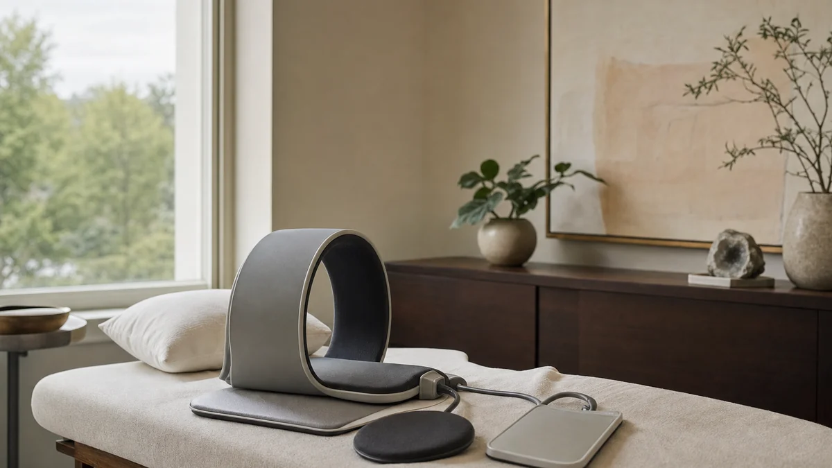
Clinical-grade PEMF devices (Physio Magnetics, BTL, MagVenture musculoskeletal systems) deliver precise, validated parameters. Field intensity, frequency, and waveform are controlled and verified. These are the devices used in most published clinical trials.
Advantages: Validated parameters, professional oversight, documented protocols for specific conditions.
Disadvantages: Cost per session, clinic access requirement, impractical for the daily sessions that most OA protocols recommend.
Home Devices
The home PEMF device market is large and heterogeneous. Devices marketed for OA vary significantly in field strength, frequency range, and waveform shape.
What to look for:
- Frequency range that includes 15–75 Hz
- Coil-style applicators (not whole-body mats for joint-specific treatment)
- Published data from the manufacturer on field intensity at the tissue level — not just “flux density at the coil surface”
What to avoid: Whole-body mat systems for joint-specific OA treatment (field too diffuse), devices that only operate at very low (sub-1 Hz) or very high (above 1,000 Hz) frequencies, and any device that lacks specifiable frequency settings.
My practical recommendation for OA: use clinical sessions for the induction phase (weeks 1–6) to establish therapeutic benefit and confirm response, then transition to a quality home device for maintenance, applying PEMF directly over the affected joint for 30–45 minutes daily.
Safety and Contraindications
PEMF has an excellent safety profile across clinical trials. Adverse events reported are rare and mild:
- Transient local warmth or mild increase in joint soreness in the first 1–2 sessions, resolving spontaneously
- Occasional dizziness with whole-body systems (not relevant for joint-specific OA application)
Absolute contraindications:
- Active electronic implants (pacemakers, cochlear implants, neurostimulators) — PEMF may interfere with device function
- Pregnancy — insufficient safety data; avoid during treatment zone
- Active malignancy in or near the treatment area
Relative contraindications / cautions:
- Acute haematoma or active bleeding at the treatment site
- Epilepsy (whole-body systems; joint-specific devices with adequate distance from the head are generally considered safe)
- Recent orthopaedic hardware in the treatment area — most titanium implants are non-magnetic and considered safe; discuss with the orthopaedic surgeon
Related Articles
- PEMF Therapy: The Complete Clinical Guide — mechanisms, general evidence, and clinical applications
- PEMF for Fibromyalgia: Evidence and Protocols — overlapping chronic pain application
- PEMF for Osteoporosis: Bone Density Evidence — adjacent musculoskeletal application
- PRP Therapy: Platelet-Rich Plasma for Joint and Tissue Healing — often combined with PEMF for OA
- Supplements for Joint Health: Collagen, Boswellia, and Omega-3 — nutritional support layer for OA protocols
References
-
Ciombor DM, Aaron RK, Wang S, Simon B. Modification of osteoarthritis by pulsed electromagnetic field — a morphological study. Osteoarthritis Cartilage. 2003;11(6):455-462. PMID: 12801487
-
Veronesi F, Torricelli P, Giavaresi G, et al. In vivo effect of two different pulsed electromagnetic field frequencies on osteoarthritis. J Orthop Res. 2014;32(5):677-685. PMID: 24395641
-
Yang X, He H, Ye W, et al. Effects of pulsed electromagnetic field therapy on pain, stiffness, physical function, and quality of life in patients with osteoarthritis: a systematic review and meta-analysis. Phys Ther. 2020;100(7):1118-1131. PMID: 32280994
-
Vavken P, Arrich F, Schuhfried O, Dorotka R. Effectiveness of pulsed electromagnetic field therapy in the management of osteoarthritis of the knee: a meta-analysis of randomized controlled trials. J Rehabil Med. 2009;41(6):406-411. PMID: 19479151
-
Battisti E, Albanese A, Guerra L, et al. Treatment with pulsed electromagnetic fields (PEMFs) in knee osteoarthritis (OA). Clin Rheumatol. 2004;23(5):386-389. PMID: 15168184
-
Thamsborg G, Florescu A, Oturai P, et al. Treatment of knee osteoarthritis with pulsed electromagnetic fields: a randomized, double-blind, placebo-controlled study. Osteoarthritis Cartilage. 2005;13(7):575-581. PMID: 15979003
-
Bagnato GL, Miceli G, Marino N, et al. Pulsed electromagnetic fields in knee osteoarthritis: a double blind, placebo-controlled, randomized clinical trial. Rheumatology (Oxford). 2016;55(4):755-762. PMID: 26705327