Lyme disease is one of the most contentious areas in medicine, and much of the controversy stems from diagnosis. The standard testing approach — a two-tier system of ELISA screening followed by Western blot confirmation — was designed to optimize specificity (minimizing false positives). The consequence is that it sacrifices sensitivity (missing true positives), particularly in chronic Lyme disease.
I treat patients with Lyme disease routinely at Klinik St. Georg, many of whom have been told they do not have Lyme disease based on negative standard testing. This does not mean their previous physicians were wrong to follow standard protocols. It means the standard protocols have documented limitations that are well-known within the scientific literature but poorly communicated in routine clinical practice.
The Standard Two-Tier System
ELISA (Enzyme-Linked Immunosorbent Assay)
The ELISA screens for antibodies (IgM and IgG) against Borrelia burgdorferi. If positive or equivocal, a Western blot is performed for confirmation.
The limitations are well-documented:
Antibody timing. ELISA detects antibodies, which take two to six weeks to develop after infection. In early Lyme disease — when treatment is most effective — up to 50% of patients test negative on ELISA because they have not yet produced sufficient antibodies. Evidence level: documented in the CDC’s own publications.
Antibody production in chronic infection. In chronic Lyme disease, some patients produce insufficient antibodies to trigger a positive ELISA. This can occur due to immune suppression by the organism, biofilm formation, intracellular sequestration, or prior antibiotic treatment that partially suppressed but did not eradicate the infection.
Strain variability. Standard ELISA kits typically use antigens from a single Borrelia strain (B31). Borrelia burgdorferi sensu lato includes multiple genospecies (B. afzelii, B. garinii, B. spielmanii in Europe) with antigenic variation. A patient infected with a strain that differs from the test antigen may test negative despite active infection.
Seronegative Lyme disease. The existence of seronegative Lyme disease — clinical Lyme disease with negative standard serology — is documented in the medical literature. It is not a fringe concept. It is a known limitation of antibody-based testing.
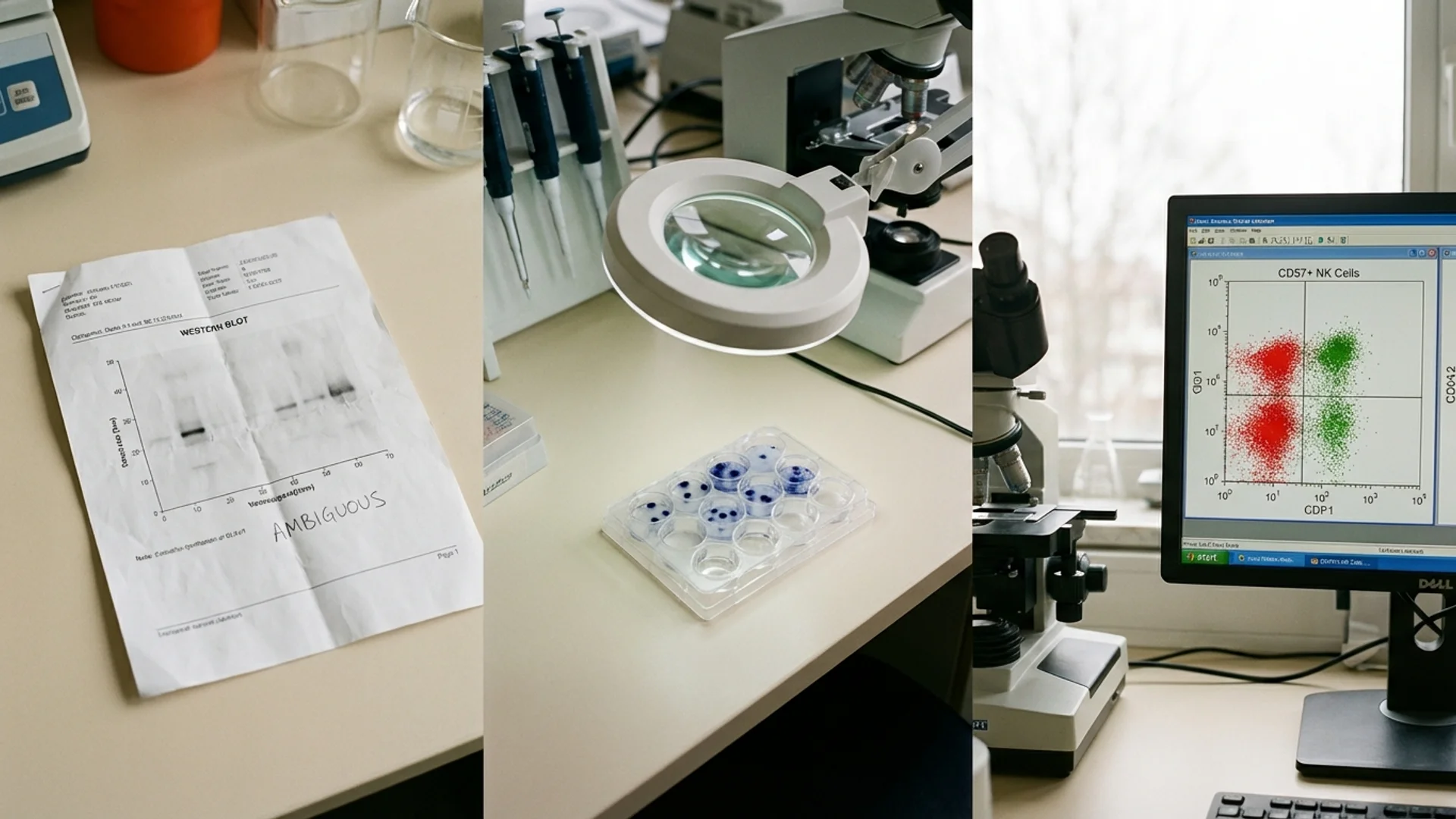
Western Blot
The Western blot identifies antibodies against specific Borrelia proteins (bands). CDC surveillance criteria require specific band patterns for positive interpretation. These criteria are appropriate for epidemiological surveillance (where specificity is prioritized) but are not necessarily appropriate for individual clinical diagnosis (where sensitivity may be more important).
Several diagnostically significant bands (OspA at 31 kDa, OspB at 34 kDa) were removed from the CDC interpretive criteria because they were used in early Lyme vaccine development. This decision was reasonable in the context of vaccine-era surveillance but reduces the test’s diagnostic sensitivity in the post-vaccine era.
Advanced Testing Approaches
ELISpot (Enzyme-Linked Immunospot)
The ELISpot assay measures cellular immune responses rather than antibody levels. It detects individual T cells that respond to Borrelia antigens by measuring cytokine (IFN-gamma) secretion at the single-cell level.
Advantages:
- Detects cellular immunity, which may persist even when antibody levels are low
- Higher analytical sensitivity than ELISA
- Can distinguish between active infection and past exposure (active T cell responses indicate ongoing antigenic stimulation)
- Not affected by the timing limitations that apply to early antibody responses
Limitations:
- Not yet widely standardized across laboratories
- Some specificity concerns (potential cross-reactivity with other spirochetes)
- Not endorsed by all infectious disease societies (though this reflects institutional conservatism as much as scientific evidence)
Evidence level: controlled studies demonstrating higher sensitivity than standard serology; clinical validation in multiple European laboratories. The ELISpot is part of standard diagnostic workup at specialized Lyme disease centers in Germany.
In my clinical experience, the ELISpot identifies Borrelia-reactive T cells in a significant proportion of patients with clinical Lyme disease who test negative on standard serology. This does not mean every negative ELISA is wrong. It means that the ELISpot provides additional diagnostic information that standard serology misses.
CD57+ NK Cells
As discussed in the NK cells article, low CD57+ NK cell counts (below 60 cells/mcL) have been observed in chronic Lyme disease and may serve as a marker of disease activity and treatment response.
I use CD57 as one data point among many. It is not specific to Lyme disease and should not be used as a standalone diagnostic test. But in the context of a clinical picture consistent with Lyme disease, a low CD57 adds supportive evidence.
Evidence level: clinical observation and retrospective studies. Further controlled research is warranted.
PCR (Polymerase Chain Reaction)
PCR detects Borrelia DNA directly. It is highly specific when positive, but sensitivity is limited by the low concentration of Borrelia in blood and most tissues. PCR from synovial fluid in Lyme arthritis has reasonable sensitivity; PCR from blood has low sensitivity (Borrelia’s blood concentration is typically very low).
Culture
Borrelia culture from patient samples is technically possible but requires specialized media, prolonged incubation (weeks to months), and has low sensitivity. It is primarily a research tool rather than a routine clinical test.
Co-Infection Testing
Tick-borne infections rarely occur in isolation. The same tick that transmits Borrelia burgdorferi can transmit:
- Babesia (babesiosis) — test with PCR and antibody panel; blood smear has low sensitivity
- Anaplasma (anaplasmosis) — PCR and antibody panel
- Ehrlichia (ehrlichiosis) — PCR and antibody panel
- Bartonella — PCR, antibody panel, and increasingly, digital PCR for improved sensitivity
- Rickettsia — antibody panel
Co-infections can alter the clinical picture significantly and may require different treatment approaches. Testing for co-infections should be standard practice in any patient being evaluated for tick-borne disease.
My Diagnostic Approach
When I evaluate a patient for suspected Lyme disease at Klinik St. Georg, the workup includes:
- Detailed clinical history — tick exposure, rash history, symptom timeline, prior treatment
- Standard serology (ELISA and Western blot) — as a baseline, understanding its limitations
- ELISpot for Borrelia-specific T cell reactivity
- Lymphocyte subset panel including CD57+ NK cells and NK cell function
- Co-infection panel (Babesia, Anaplasma, Bartonella, Rickettsia)
- Inflammatory markers (CRP, cytokines) to assess current inflammatory burden
- Comprehensive metabolic assessment to identify complicating factors
No single test is definitive for Lyme disease. The diagnosis is clinical, supported by laboratory findings. A patient with classic symptoms, documented tick exposure, positive ELISpot, low CD57, and negative standard serology has Lyme disease — regardless of what the ELISA says.
What I tell my patients: Lyme disease testing is imperfect. The standard tests miss cases. The advanced tests are not perfect either. The best diagnostic approach combines multiple testing modalities with careful clinical assessment. Any physician who relies solely on ELISA results to rule out Lyme disease is applying an incomplete diagnostic standard.
Disclaimer: This article is provided for educational purposes and reflects one physician’s clinical perspective. Lyme disease diagnosis and treatment remain areas of active scientific debate. Consult a physician experienced in tick-borne diseases for diagnostic and treatment decisions.