At a Glance
| Property | Value |
|---|---|
| Evidence Level | Moderate |
| Primary Use | Understanding the multi-viral reactivation cascade in chronic illness |
| Key Mechanism | Reactivated herpesviruses produce immunomodulatory proteins that suppress NK cell and T-cell function, enabling further viral reactivation |
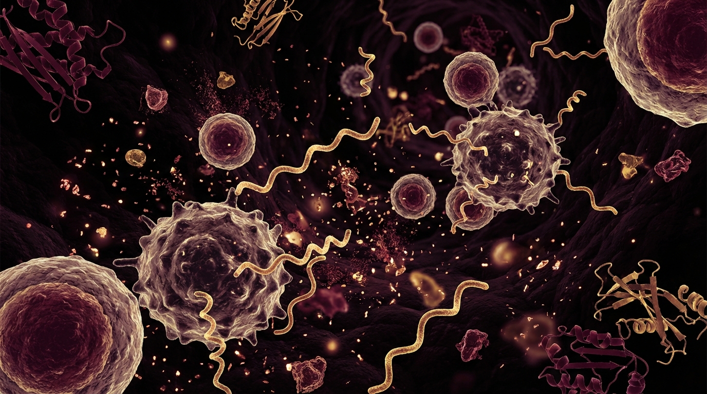
The Vicious Cycle Nobody Explains
Here is a question I hear constantly in clinical practice: “How did I end up with three reactivated viruses? I thought my only problem was Lyme disease.”
The answer reveals one of the most important mechanisms in chronic infectious disease — and one that is rarely explained to patients clearly. Reactivated viruses are not passive passengers. They are active saboteurs of the immune system that should be controlling them.
Let me be direct about what this means: each reactivated virus makes it easier for the next one to reactivate. EBV suppresses the NK cells that should control HHV-6. HHV-6 suppresses the T-cells that should control CMV. CMV exhausts the immune repertoire that should control all of them. The result is a cascading failure of viral containment that compounds itself — and that explains why patients with chronic infections so rarely have just one reactivated virus.
The Three Virus Players
EBV (Epstein-Barr Virus / HHV-4)
Immune suppression mechanisms:
Viral IL-10 (vIL-10): EBV produces a protein called vIL-10 that is structurally similar to human IL-10 — a potent immunosuppressive cytokine. vIL-10 suppresses T-cell proliferation, reduces NK cell cytotoxicity, and inhibits the production of pro-inflammatory cytokines (TNF-alpha, IL-12) that are essential for anti-viral immune responses [1].
In practical terms: EBV produces its own immunosuppressive signal that mimics your body’s natural “stand down” command. Your immune cells receive a signal that looks legitimate and reduce their surveillance — allowing EBV to persist and other viruses to reactivate.
EBNA1 immune evasion: EBV nuclear antigen 1 (EBNA1) contains a glycine-alanine repeat domain that prevents its own proteasomal degradation and MHC class I presentation. This means infected cells cannot display EBV antigens to CD8+ cytotoxic T-cells — the primary killers of virus-infected cells.
BCRF1 and BARF1: Additional EBV proteins that interfere with interferon signaling and apoptosis of infected cells.
HHV-6 (Human Herpesvirus 6)
Immune suppression mechanisms:
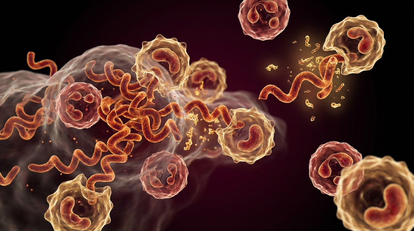
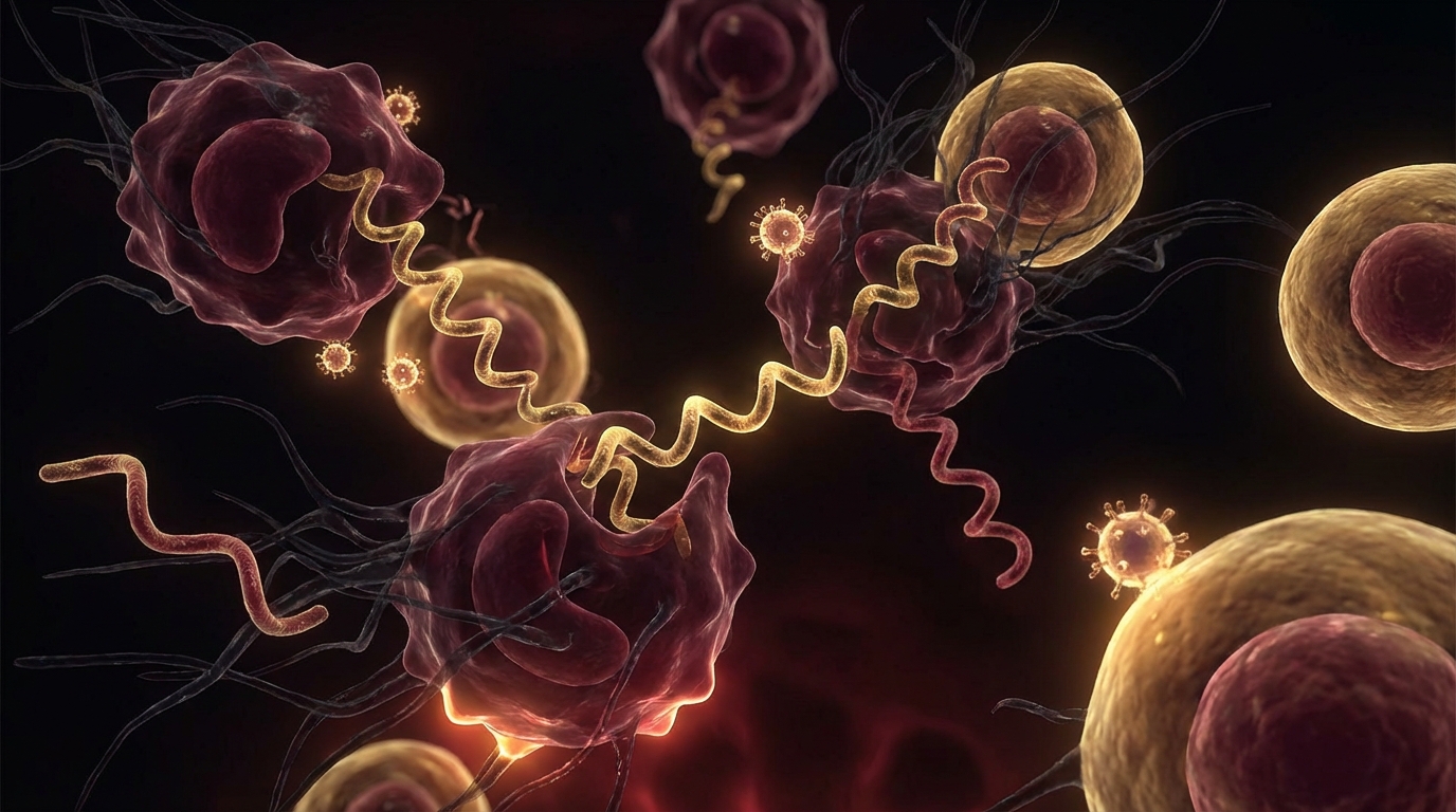
Direct NK cell infection: HHV-6 can directly infect and kill NK cells — the first line of defense against virus-infected cells. A study in Blood demonstrated that HHV-6 infection reduced NK cell cytotoxicity by up to 80% in vitro [2]. This is not subtle immune modulation; it is direct destruction of the immune cells responsible for viral surveillance.
CD4+ T-cell infection: HHV-6 infects CD4+ helper T-cells (using CD46 as its receptor), disrupting the coordination of adaptive immune responses. This is mechanistically similar to HIV’s CD4 tropism, though less aggressive.
Cytokine modulation: HHV-6 alters the cytokine environment, shifting from Th1 (anti-viral) to Th2 (allergic/humoral) immune profiles. This Th1/Th2 shift impairs cell-mediated immunity while potentially contributing to allergic and autoimmune manifestations.
Complement evasion: HHV-6 encodes proteins that bind complement regulatory proteins, preventing complement-mediated destruction of infected cells.
CMV (Cytomegalovirus / HHV-5)
Immune suppression mechanisms:
T-cell exhaustion and repertoire inflation: CMV is perhaps the most insidious of the three in its long-term immune effects. Over time, the immune system dedicates an increasingly large proportion of its T-cell repertoire to CMV surveillance — a phenomenon called memory inflation. In elderly individuals, up to 30-40% of the total CD8+ T-cell population may be CMV-specific [3].
This massive dedication of immune resources to CMV leaves fewer T-cells available for surveillance of other threats — including other reactivated viruses, cancer cells, and bacterial infections.
MHC class I downregulation: CMV produces multiple proteins (US2, US3, US6, US11) that interfere with MHC class I surface expression on infected cells, making them invisible to CD8+ cytotoxic T-cells.
NK cell receptor manipulation: CMV produces a viral UL16-binding protein that interferes with NKG2D — one of the primary activating receptors on NK cells. This directly impairs NK cell-mediated killing of infected cells.
Viral chemokine decoys: CMV produces proteins that mimic human chemokines, misdirecting immune cells away from sites of active infection.

The Cascade in Action
Stage 1: The Initial Trigger
A previously healthy individual develops a chronic infection — Lyme disease, in our clinical context — or experiences prolonged immune stress (mold exposure, chronic stress, surgery, COVID-19).
The initial trigger suppresses:
- NK cell function (reduced CD56bright and CD56dim subsets)
- CD8+ T-cell cytotoxicity
- Interferon-gamma production
Stage 2: First Virus Reactivates
With reduced surveillance, the most sensitive latent virus reactivates. In most patients, this is EBV — because EBV latency is maintained by the narrowest immune margin (it lives in B-cells and requires constant T-cell and NK cell surveillance).
EBV reactivation produces:
- Fatigue, cognitive dysfunction, sore throat
- vIL-10 production → further immune suppression
- NK cell function drops further
Stage 3: Second Virus Reactivates
The additional immune suppression from EBV reactivation allows HHV-6 to reactivate. HHV-6 infects and destroys NK cells, creating an even deeper immune deficit.
HHV-6 reactivation produces:
- Neurological symptoms (brain fog, neuropathy, cognitive decline)
- Direct NK cell depletion
- CD4+ T-cell dysfunction
Stage 4: Full Cascade
With both EBV and HHV-6 actively suppressing immunity, CMV may reactivate. T-cell repertoire begins to inflate toward CMV-specific responses. Other latent viruses (VZV — causing shingles outbreaks, HSV — causing cold sore recurrences) may also reactivate.
At this stage, the patient’s immune system is fighting on multiple viral fronts simultaneously while also trying to clear the original trigger (Lyme, mold). The total infectious burden overwhelms available immune resources.
The Evidence
What We Know (Human Data)
The 72.5% co-expression finding: A 2023 study published in Journal of Translational Medicine analyzed herpesvirus co-reactivation in ME/CFS patients and found that 72.5% had simultaneous active infection with multiple herpesviruses — most commonly EBV and HHV-6 together [4]. This co-reactivation rate was dramatically higher than in healthy controls.
NK cell dysfunction in chronic Lyme: Multiple studies have documented reduced NK cell function in chronic Lyme patients. A study in Frontiers in Immunology showed that ME/CFS patients (many with tick-borne illness history) had significantly reduced NK cell cytotoxicity compared to controls — the same immune deficit that enables viral reactivation [5].
The immune suppression mechanisms described above are individually well-characterized. vIL-10 production by EBV, NK cell infection by HHV-6, and T-cell repertoire inflation by CMV are documented in virology literature. What is newer is the clinical framework connecting these mechanisms into a cascade model in chronic infection patients.
What I See in Practice
In our clinical experience, the cascade model explains patterns we observe daily:
Pattern 1: Sequential reactivation. Patients who develop Lyme disease often present first with bacterial symptoms, then develop fatigue and cognitive decline disproportionate to their Lyme burden — corresponding to viral reactivation occurring weeks to months after immune suppression from the primary infection.
Pattern 2: Treatment plateau. Patients improve partially with anti-Borrelia therapy but hit a plateau. When viral reactivation is identified and treated, a second phase of improvement occurs. This two-phase recovery pattern is consistent with addressing the bacterial trigger and then the viral secondary infections.
Pattern 3: Treatment-resistant cases. The most severe and treatment-resistant patients almost always have the highest viral co-reactivation burden. In my clinical experience, patients with three or more reactivated viruses (EBV + HHV-6 + CMV or VZV) are the most challenging to treat and require the longest treatment durations.
What I tell my patients: your immune system is fighting a war on multiple fronts. Each front it loses ground on makes every other front harder. Treatment must address all active infections and restore immune function — not just target one pathogen at a time.
Practical Application
Comprehensive Viral Assessment
For any chronic infection patient with disproportionate fatigue, neurological symptoms, or treatment plateau, the following panel is recommended:
EBV:
- VCA IgM, VCA IgG, EA IgG, EBNA IgG
- EBV quantitative PCR (if serology suggests reactivation)
HHV-6:
- HHV-6 IgG (baseline — present in 90%+)
- HHV-6 quantitative PCR (active replication)
- HHV-6 IgG avidity (recent vs. remote infection)
CMV:
- CMV IgM, CMV IgG
- CMV quantitative PCR (if IgM positive or symptoms suggestive)
Immune function:
- NK cell function (CD56 bright and dim subsets, cytotoxicity assay)
- Lymphocyte subset panel (CD4, CD8, CD4/CD8 ratio)
- CD57 NK cell count (though interpretation is debated)
Breaking the Cycle: Treatment Strategy
Step 1: Identify and treat the primary trigger
- Continue Lyme/co-infection treatment
- Address mold/CIRS if present
- Optimize sleep, stress, and nutrition
Step 2: Targeted antiviral therapy
- EBV: Valacyclovir 1g TID
- HHV-6: Valganciclovir 450-900mg BID
- CMV: Valganciclovir (same agent treats both HHV-6 and CMV)
Step 3: Immune restoration
- Thymosin alpha-1 — T-cell and NK cell enhancement
- Vitamin D optimization — 50-80 ng/mL
- Zinc 30-50mg daily
- Medicinal mushrooms (AHCC, turkey tail, reishi)
- Low-dose naltrexone (4.5mg nightly) — evidence for NK cell upregulation
Step 4: Monitor viral loads and immune markers
- PCR for each reactivated virus every 3 months
- NK cell function testing every 3-6 months
- Adjust therapy based on viral clearance and immune recovery

Safety and Considerations
- Valganciclovir requires regular CBC monitoring due to bone marrow suppression risk. Do not initiate without a monitoring plan.
- Treating multiple viral infections simultaneously is complex and requires experienced supervision. Drug interactions, cumulative toxicity, and Herxheimer-like reactions must be managed.
- Low-dose naltrexone (LDN) is used off-label for immune modulation. It is generally well-tolerated but can interfere with opioid medications. Do not use in patients taking opioid analgesics.
- The immune restoration phase takes time. Expect months, not weeks, for measurable improvement in NK cell function and viral load reduction.
- Not all patients need antiviral therapy. If viral reactivation markers are borderline and immune function is intact, supporting the immune system while treating the primary infection may be sufficient.
The Bottom Line
Reactivated viruses are not passive bystanders in chronic illness — they are active participants in immune suppression. EBV, HHV-6, and CMV each employ sophisticated strategies to evade and suppress the immune responses that should keep them dormant. When multiple viruses reactivate simultaneously — as they do in the majority of chronic infection patients — the cumulative immune suppression creates a self-perpetuating cycle that cannot be broken by treating any single component alone. Here is what the evidence shows: comprehensive viral assessment, targeted antiviral therapy, and deliberate immune restoration are required to break the cascade. In my clinical experience, patients who address the full viral picture — not just the primary bacterial infection — achieve deeper and more durable recovery.
References
- Jochum S, Ruiss R, Moosmann A, Hammerschmidt W, Zeidler R. RNAs in Epstein-Barr virions control early steps of infection. Proc Natl Acad Sci U S A. 2012;109(21):E1396-E1404. PMID: 22543160
- Flamand L, Stefanescu I, Menezes J. Human herpesvirus-6 enhances natural killer cell cytotoxicity via IL-15. J Clin Invest. 1996;97(6):1373-1381. PMID: 8617868
- Klenerman P, Oxenius A. T cell responses to cytomegalovirus. Nat Rev Immunol. 2016;16(6):367-377. PMID: 27108521
- Rasa-Dzelzkaleja S, Krumina A, Capenko S, et al. The persistent viral infections in the development and severity of myalgic encephalomyelitis/chronic fatigue syndrome. J Transl Med. 2023;21(1):33. PMID: 36653843
- Brenu EW, van Driel ML, Staines DR, et al. Immunological abnormalities as potential biomarkers in chronic fatigue syndrome/myalgic encephalomyelitis. J Transl Med. 2011;9:81. PMID: 21619669