At a Glance
| Property | Value |
|---|---|
| Evidence Level | Strong (individual biomarkers); Clinical observation (optimal range interpretation) |
| Primary Use | Comprehensive thyroid function assessment and Hashimoto’s screening |
| Key Concept | TSH alone is insufficient; conversion, autoimmunity, and cellular uptake require additional markers |
Why TSH Alone Is Not Enough
If you have ever been told your thyroid is “fine” based on a single TSH test, while you sit there feeling exhausted, cold, foggy, and ten kilograms heavier than you should be, this article is for you.
TSH (thyroid-stimulating hormone) is the most commonly ordered thyroid test in the world. It is also, when used in isolation, one of the most misleading tests in endocrinology. Not because the test itself is flawed — TSH is a well-validated, highly sensitive assay — but because thyroid physiology is a multi-step cascade, and TSH only measures the first step.
Here’s what the research actually says about how thyroid hormone works in the body, why TSH alone misses important clinical information, and what a complete panel looks like.
The Thyroid Cascade: From Brain to Cell
Understanding why you need more than TSH requires understanding how thyroid hormones get from your brain to your cells. This is not optional complexity — each step can malfunction independently, and each requires a different test to evaluate.
Step 1: The Brain Signals the Thyroid (TSH)
The hypothalamus releases TRH (thyrotropin-releasing hormone), which signals the pituitary gland to release TSH. TSH then travels to the thyroid gland and stimulates production of thyroid hormones. When thyroid hormone levels are adequate, the pituitary reduces TSH output (negative feedback). When levels are low, TSH rises.
This is the step that TSH measures: the pituitary gland’s perception of circulating thyroid hormone. If TSH is high, the pituitary thinks there is not enough thyroid hormone. If TSH is low, it thinks there is too much.
The limitation: TSH tells you what the pituitary thinks. It does not tell you what is happening at the cellular level in the liver, brain, muscles, or gut.
Step 2: The Thyroid Produces T4 (Measured by Free T4)
The thyroid gland produces predominantly T4 (thyroxine) — approximately 80-90% T4 and 10-20% T3. T4 is a prohormone: it is relatively inactive and must be converted to T3 (the active form) to exert metabolic effects.
Free T4 measures the unbound, bioavailable fraction of T4 in the bloodstream. Low free T4 with elevated TSH confirms primary hypothyroidism. Normal free T4 with elevated TSH suggests subclinical hypothyroidism — the thyroid is still maintaining output, but only because the pituitary is pushing harder.
Step 3: T4 Converts to T3 (Measured by Free T3)
The conversion of T4 to T3 occurs primarily in the liver, kidneys, and peripheral tissues via deiodinase enzymes (type 1 and type 2). This is the critical activation step — without adequate conversion, a patient can have normal TSH and normal free T4 but functionally low T3 at the cellular level.
Factors that impair T4-to-T3 conversion:
- Chronic stress and elevated cortisol — directly inhibit deiodinase enzymes
- Selenium deficiency — deiodinase enzymes are selenoproteins
- Zinc deficiency — required cofactor for conversion
- Iron deficiency — impairs thyroid peroxidase activity and conversion
- Chronic inflammation — inflammatory cytokines (IL-6, TNF-alpha) suppress deiodinase activity
- Caloric restriction and crash dieting — the body downregulates T3 production to conserve energy
- Liver dysfunction — impaired hepatic conversion
- Gut dysbiosis — approximately 20% of T4-to-T3 conversion occurs in the gut, mediated by intestinal sulfatase-producing bacteria
- Certain medications — amiodarone, beta-blockers, lithium, corticosteroids
In my clinical experience, impaired T4-to-T3 conversion is one of the most common and most overlooked causes of persistent hypothyroid symptoms despite “normal” TSH and “normal” T4. Free T3 is the only way to identify it.
Step 4: Reverse T3 — The Brake Pedal
When the body converts T4 to reverse T3 (rT3) instead of active T3, it is applying the metabolic brake. Reverse T3 is biologically inactive — it binds to thyroid receptors but does not activate them, effectively blocking the action of T3 at the cellular level.
Reverse T3 rises in:
- Chronic illness (“sick euthyroid syndrome” or “non-thyroidal illness”)
- Chronic stress and high cortisol
- Caloric restriction
- Inflammation
- Surgery and trauma
- Selenium deficiency
A patient with normal TSH, normal free T4, low-normal free T3, and elevated reverse T3 has a conversion problem that TSH alone completely misses. The ratio of free T3 to reverse T3 is clinically informative — a ratio below 0.2 (when T3 is measured in pg/mL and rT3 in ng/dL) suggests excessive reverse T3 dominance.

Step 5: Autoimmune Attack (Measured by TPO and TG Antibodies)
Hashimoto’s thyroiditis is the most common cause of hypothyroidism in iodine-sufficient countries, affecting an estimated 5-10% of the general population and up to 20% of women over 60 [1]. It is an autoimmune condition in which the immune system attacks the thyroid gland, gradually destroying its ability to produce hormones.
The critical clinical fact: thyroid antibodies (TPO-Ab and TG-Ab) are often elevated for years to decades before TSH becomes abnormal. A patient can have active autoimmune thyroid destruction with a completely “normal” TSH because the remaining thyroid tissue is still compensating — for now.
TPO Antibodies (Anti-thyroid peroxidase): The most sensitive marker for Hashimoto’s. Elevated in approximately 90% of Hashimoto’s cases. Thyroid peroxidase is the enzyme that catalyzes iodine organification and thyroid hormone synthesis — antibodies against it directly impair hormone production.
TG Antibodies (Anti-thyroglobulin): Elevated in approximately 60-70% of Hashimoto’s cases. Sometimes the only elevated antibody in early or mild autoimmune thyroiditis. Also relevant in thyroid cancer monitoring (TG-Ab can interfere with thyroglobulin assays).
What I tell my patients: if I only test TSH and find it normal, I might send home a patient whose immune system is actively destroying their thyroid gland. Testing antibodies catches Hashimoto’s at its earliest stage — when lifestyle intervention, selenium supplementation, and targeted immune modulation can potentially slow the autoimmune process before irreversible thyroid damage occurs.
The Complete Panel: What to Order
| Test | What It Measures | Optimal Range | Standard Range |
|---|---|---|---|
| TSH | Pituitary signal to thyroid | 0.5 - 2.0 mIU/L | 0.4 - 4.5 mIU/L |
| Free T4 | Available prohormone | 1.1 - 1.5 ng/dL (upper half of range) | 0.8 - 1.8 ng/dL |
| Free T3 | Active thyroid hormone | 3.0 - 4.0 pg/mL (upper third of range) | 2.3 - 4.2 pg/mL |
| Reverse T3 | Inactive T3 (metabolic brake) | < 15 ng/dL | 8 - 25 ng/dL |
| Free T3:rT3 ratio | Conversion efficiency | > 0.2 | Not routinely calculated |
| TPO Antibodies | Autoimmune thyroiditis marker | < 35 IU/mL (ideally < 20) | < 35 IU/mL |
| TG Antibodies | Autoimmune thyroiditis marker | < 20 IU/mL | < 40 IU/mL (varies by lab) |
I also recommend checking these alongside the thyroid panel, as they directly affect thyroid function:
| Supporting Test | Why |
|---|---|
| Ferritin | Iron is required for thyroid peroxidase activity |
| Selenium (or selenoprotein P) | Deiodinase enzymes are selenoproteins |
| Vitamin D (25-OH-D) | Low vitamin D is associated with higher Hashimoto’s prevalence |
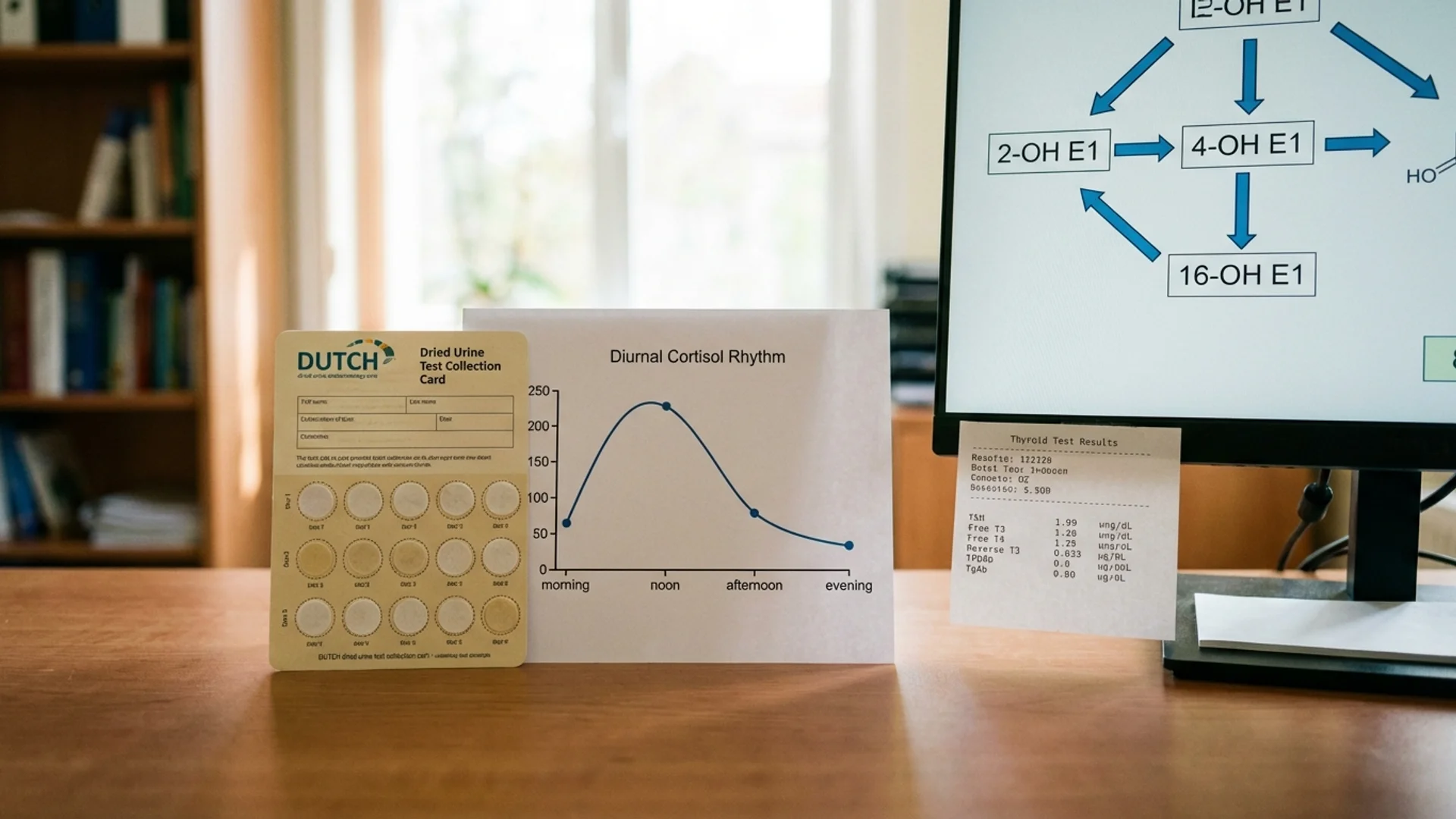
| Cortisol (AM or DUTCH test) | Elevated cortisol impairs T4-to-T3 conversion |
For more on optimal ranges for these supporting markers, see my optimal lab ranges article.
How to Interpret Common Patterns
Pattern 1: Classic Primary Hypothyroidism
- TSH: elevated (> 4.5)
- Free T4: low or low-normal
- Free T3: low
- Antibodies: may or may not be elevated
This is the textbook pattern. Most doctors will catch this. Treatment is typically levothyroxine (synthetic T4).
Pattern 2: Subclinical Hypothyroidism
- TSH: mildly elevated (2.5 - 4.5) or upper-normal
- Free T4: normal
- Free T3: normal or low-normal
- Antibodies: often positive
This is the grey zone that generates the most clinical disagreement. Standard endocrinology guidelines often recommend “watch and wait” until TSH exceeds 10. Functional and integrative physicians — myself included — believe that symptomatic patients in this range deserve a trial of treatment, particularly if antibodies are positive.
The evidence supports intervention: a 2019 meta-analysis showed that treatment of subclinical hypothyroidism improves lipid profiles, cardiac function, and quality of life, particularly in patients under 65 with TSH above 7 [2]. Below 7, the data is less clear, and the clinical decision depends heavily on symptoms, antibody status, and clinical context.
Pattern 3: Conversion Problem (Low T3 Syndrome)
- TSH: normal (often 1.0 - 2.5)
- Free T4: normal or high-normal
- Free T3: low or low-normal
- Reverse T3: elevated
- Free T3:rT3 ratio: < 0.2
This pattern is invisible on standard TSH-only testing. The pituitary sees adequate T4 and keeps TSH normal, but the body is not converting T4 to active T3. The patient has hypothyroid symptoms with “normal” labs.
Causes include chronic stress, inflammation, selenium or iron deficiency, caloric restriction, and chronic illness. Treatment focuses on addressing the underlying conversion block (stress reduction, nutritional repletion, inflammation management) rather than simply adding more T4.
In some cases, I prescribe combination T4/T3 therapy or desiccated thyroid (which contains both T4 and T3) for patients with documented conversion problems who do not respond to T4 monotherapy. This remains controversial in mainstream endocrinology but is well-supported by clinical experience and several RCTs showing patient preference and symptom improvement with combination therapy [3].
Pattern 4: Early Hashimoto’s (Euthyroid Autoimmune Thyroiditis)
- TSH: normal
- Free T4: normal
- Free T3: normal
- TPO Antibodies: elevated (often > 100 IU/mL)
- TG Antibodies: may be elevated
This patient has normal thyroid function — for now. But the immune system is attacking the thyroid, and over the next 5-20 years, function will progressively decline. Identifying this pattern early opens a window for immune modulation, gluten elimination (there is a well-documented association between celiac disease/gluten sensitivity and Hashimoto’s [4]), selenium supplementation (200 mcg/day reduces TPO antibodies by approximately 20-40% in multiple RCTs [5]), and lifestyle optimization.
This is the pattern that justifies routine antibody testing even when TSH is normal.
Pattern 5: Central (Pituitary) Hypothyroidism
- TSH: normal or low-normal (inappropriately normal given low thyroid hormones)
- Free T4: low
- Free T3: low
This rare but important pattern occurs when the pituitary gland fails to produce adequate TSH despite low thyroid hormone levels. Causes include pituitary adenomas, post-surgical pituitary damage, traumatic brain injury, and chronic high-dose glucocorticoid use. TSH-only testing will completely miss this — the TSH looks “normal” while the patient is profoundly hypothyroid.

Hashimoto’s Screening: Who Should Be Tested
I recommend thyroid antibody testing in:
- All women over 35 — Hashimoto’s prevalence increases sharply after 35 and peaks in the perimenopausal and postmenopausal years
- Any patient with hypothyroid symptoms regardless of TSH — fatigue, weight gain, cold intolerance, constipation, hair loss, dry skin, depression, brain fog
- Patients with other autoimmune conditions — type 1 diabetes, celiac disease, rheumatoid arthritis, vitiligo, pernicious anemia (autoimmune conditions cluster)
- Family history of thyroid disease — first-degree relatives of Hashimoto’s patients have a 5-10x increased risk
- Postpartum women — postpartum thyroiditis affects 5-10% of women and is often Hashimoto’s presenting for the first time
- Patients with infertility or recurrent miscarriage — thyroid antibodies are independently associated with reproductive complications even when TSH is normal [6]
- Patients with elevated cholesterol resistant to treatment — hypothyroidism is an underdiagnosed cause of hyperlipidemia
What I See in Practice
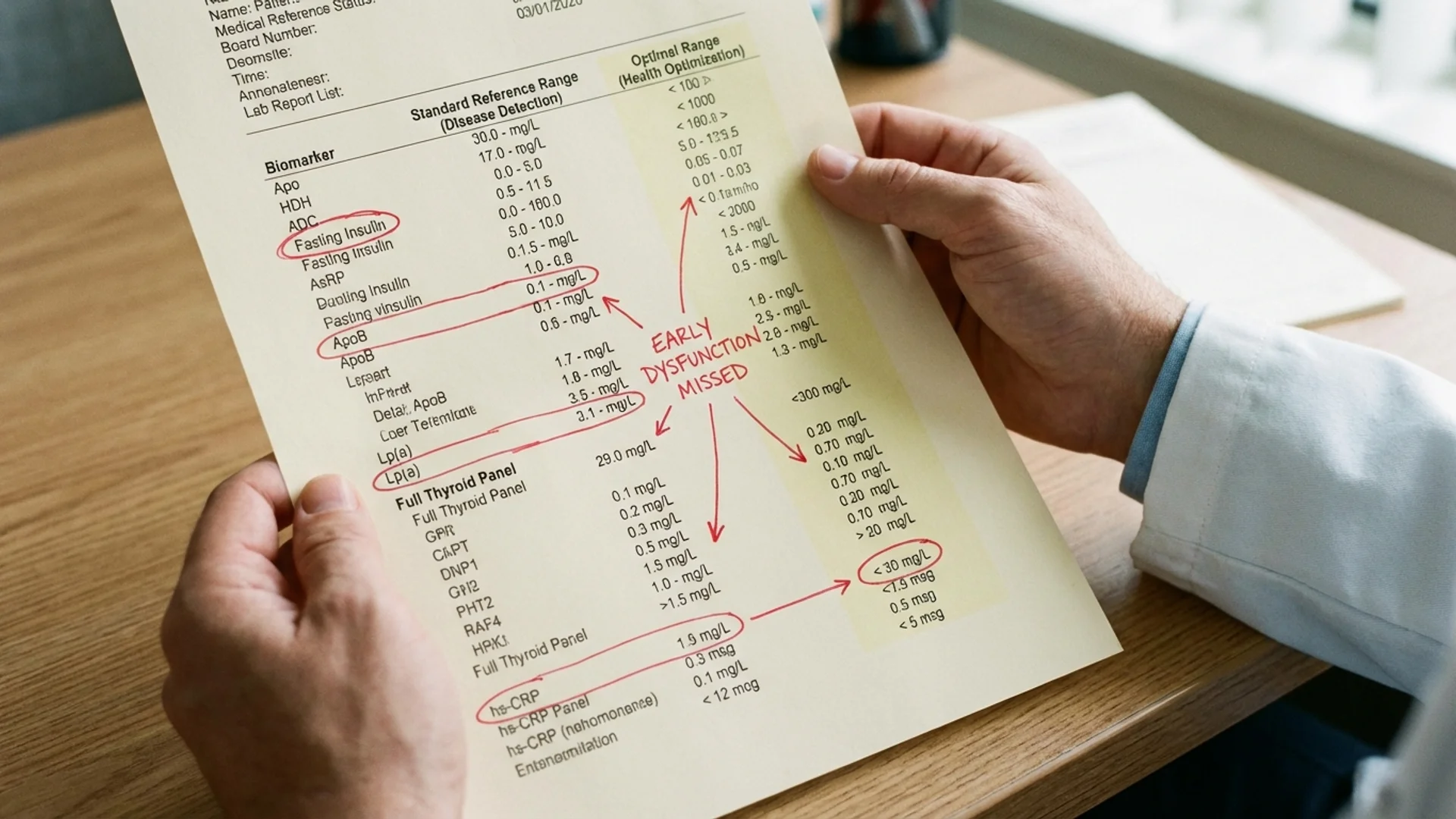
In our clinical experience treating patients from over 90 countries, thyroid dysfunction is one of the most commonly missed diagnoses — not because the testing is unavailable, but because it is incompletely performed. I estimate that 30-40% of patients we see with chronic fatigue, unexplained weight gain, or treatment-resistant depression have a thyroid component that was missed by TSH-only testing.
The most common scenario: a woman in her late 30s to early 50s, progressively fatigued, gaining weight despite exercise, losing hair, and feeling cognitively dull. Multiple doctors have checked TSH, found it between 2.0 and 4.0, and told her she is “fine.” A complete panel reveals elevated TPO antibodies (often > 200 IU/mL), a low free T3, an elevated reverse T3, and iron deficiency — a constellation of findings that explains her symptoms and is eminently treatable.
This is not rare. This is routine. And it is missed because of an overreliance on a single screening test.
Safety and Considerations
- Do not self-diagnose or self-treat thyroid conditions. Thyroid hormone medication requires physician supervision, dosing titration, and monitoring.
- Thyroid antibody levels fluctuate. A single elevated result confirms autoimmune thyroiditis, but the antibody titer does not correlate linearly with disease severity. Trending antibodies over time (every 6-12 months) is more informative than a single measurement.
- Biotin supplementation interferes with thyroid assays. If you take biotin (a common supplement in hair/skin products), discontinue it 48-72 hours before thyroid blood work. Biotin causes falsely low TSH and falsely high free T4/T3 on certain immunoassay platforms, which can lead to misdiagnosis of hyperthyroidism.
- Time of testing matters. TSH has a circadian rhythm, peaking in the early morning and reaching a nadir in the afternoon. For consistent results, draw thyroid panels in the morning (before 10 AM), fasting if possible.
- Levothyroxine timing. If you take thyroid medication, do not take it the morning of your blood draw — this will produce a falsely elevated free T4. Take it after the blood draw.
The Bottom Line
A complete thyroid panel — TSH, free T3, free T4, reverse T3, TPO antibodies, and TG antibodies — is the minimum required to adequately assess thyroid function. TSH alone is a screening test that misses conversion problems, early autoimmune thyroiditis, reverse T3 dominance, and central hypothyroidism. If you have symptoms consistent with thyroid dysfunction and your doctor has only checked TSH, you do not yet know whether your thyroid is functioning properly. Request the full panel. The nuance matters.
References
- Vanderpump MP. The epidemiology of thyroid disease. Br Med Bull. 2011;99:39-51. PMID: 21893493
- Feller M, Snel M, Moutzouri E, et al. Association of thyroid hormone therapy with quality of life and thyroid-related symptoms in patients with subclinical hypothyroidism: a systematic review and meta-analysis. JAMA. 2018;320(13):1349-1359. PMID: 30285179
- Wiersinga WM, Duntas L, Fadeyev V, Nygaard B, Vanderpump MP. 2012 ETA guidelines: the use of L-T4 + L-T3 in the treatment of hypothyroidism. Eur Thyroid J. 2012;1(2):55-71. PMID: 24782999
- Sategna-Guidetti C, Volta U, Ciacci C, et al. Prevalence of thyroid disorders in untreated adult celiac disease patients and effect of gluten withdrawal: an Italian multicenter study. Am J Gastroenterol. 2001;96(3):751-757. PMID: 11280546
- Wichman J, Winther KH, Bonnema SJ, Hegedus L. Selenium supplementation significantly reduces thyroid autoantibody levels in patients with chronic autoimmune thyroiditis: a systematic review and meta-analysis. Thyroid. 2016;26(12):1681-1692. PMID: 27702392
- Thangaratinam S, Tan A, Knox E, Kilby MD, Franklyn J, Coomarasamy A. Association between thyroid autoantibodies and miscarriage and preterm birth: meta-analysis of evidence. BMJ. 2011;342:d2616. PMID: 21558126
- Jonklaas J, Bianco AC, Bauer AJ, et al. Guidelines for the treatment of hypothyroidism: prepared by the American Thyroid Association task force on thyroid hormone replacement. Thyroid. 2014;24(12):1670-1751. PMID: 25266247