At a Glance
| Property | Value |
|---|---|
| Evidence Level | Strong (well-validated flow cytometry, established clinical utility) |
| Primary Use | Characterizing immune cell populations in chronic illness |
| Key Mechanism | Flow cytometry counts and classifies lymphocyte populations using surface marker antibodies |
What the Panel Measures
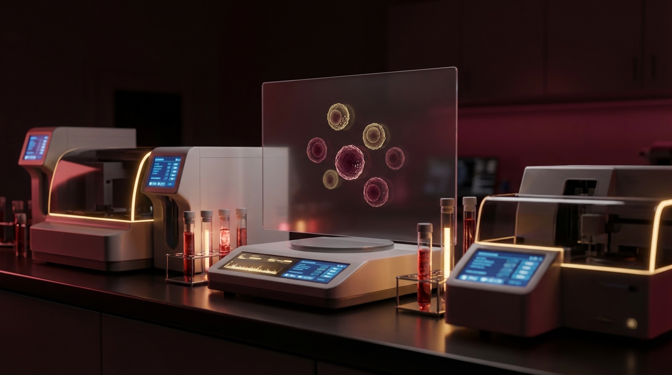
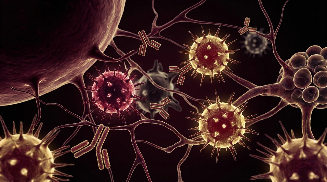
A lymphocyte subset panel — also called a lymphocyte immunophenotyping panel or T/B/NK cell panel — uses flow cytometry to identify and count the major populations of lymphocytes in your blood based on the cluster of differentiation (CD) markers on their surface.
Think of it as a detailed census of your immune army. A CBC (complete blood count) tells you the total number of lymphocytes. A lymphocyte subset panel tells you how many of each type you have and what proportions they exist in.
The standard panel reports:
CD3+ T Cells (Total T Cells)
All mature T cells express CD3. This is the total T cell count. T cells are the backbone of the adaptive immune system — they coordinate immune responses and directly kill infected cells.
Normal range: 700-2,100 cells/uL (approximately 60-80% of lymphocytes)
CD4+ T Helper Cells
CD4+ T cells orchestrate the immune response. They do not kill targets directly but activate other immune cells — B cells, macrophages, and CD8+ T cells — through cytokine signaling. They are the “generals” of the immune system.
Normal range: 500-1,500 cells/uL (30-60% of T cells)
Subtypes to be aware of: CD4+ T cells include Th1 (drive intracellular pathogen killing), Th2 (drive antibody responses and allergic reactions), Th17 (drive mucosal immunity and autoimmunity), and Treg (regulatory T cells that suppress excessive immune responses). Standard panels do not differentiate these subtypes, but some advanced panels do.
CD8+ Cytotoxic T Cells
CD8+ T cells are the “special forces” — they directly kill virus-infected cells and tumor cells that present foreign antigens on MHC class I. They are critical for controlling intracellular infections [1].
Normal range: 200-900 cells/uL (15-40% of T cells)
CD4/CD8 Ratio
The ratio of helper to cytotoxic T cells is one of the most clinically useful parameters on the panel.
Normal range: 1.0-3.5
- Low ratio (<1.0, “inverted”): Suggests chronic viral activation, persistent immune stimulation. Seen in HIV (the classic example), chronic EBV/CMV reactivation, and some chronic Lyme presentations. The immune system is expanding its cytotoxic arm at the expense of its helper arm.
- High ratio (>3.5): Can indicate immune exhaustion, autoimmune tendencies, or early HIV seroconversion (paradoxically).
CD19+ B Cells
B cells produce antibodies. They are the humoral immune arm — responsible for producing the immunoglobulins (IgG, IgM, IgA) that target extracellular pathogens and toxins.
Normal range: 100-500 cells/uL (5-20% of lymphocytes)
CD56+ NK Cells
Natural killer cells — innate immune cells that kill virus-infected and malignant cells without prior sensitization.
Normal range: 90-600 cells/uL (5-20% of lymphocytes)
Note: NK cell count from a subset panel tells you how many NK cells you have. It does not tell you how well they function. NK cell function testing requires a separate cytotoxicity assay.
Common Patterns in Chronic Illness
The real value of the lymphocyte subset panel is not in isolated numbers but in the patterns they form. Here is what I commonly see in specific conditions:
Chronic Lyme Disease
- CD4/CD8 ratio: Often inverted or low-normal (chronic T cell activation)
- CD57+ NK cells: Frequently reduced (Stricker’s finding — see CD57 in Lyme)
- CD8+ T cells: May be elevated (expanded cytotoxic response to persistent intracellular infection)
- Total lymphocyte count: Variable — can be low, normal, or even elevated
ME/CFS (Chronic Fatigue Syndrome)
- NK cells: Consistently reduced in count and/or function
- CD8+ T cells: Often elevated, with increased expression of activation markers
- CD4/CD8 ratio: Frequently low or inverted
- T cell activation markers (HLA-DR, CD38): Often elevated — reflecting ongoing immune activation without resolution
Post-COVID / Long COVID
- CD4+ T cells: Often reduced in the acute phase; may not fully recover
- CD8+ T cells: Expanded, often with exhaustion markers (PD-1, Tim-3)
- NK cells: Frequently reduced in function
- B cells: Sometimes showing abnormal activation patterns
Autoimmune Conditions
- CD4/CD8 ratio: Often elevated (helper T cell-driven process)
- Treg cells: May be reduced (loss of immune regulation)
- B cells: May be elevated (autoantibody production)

The Evidence
What We Know (Human Data)
Lymphocyte subset analysis has been a clinical staple since the HIV epidemic, where CD4 count became the primary marker for disease staging and treatment decisions. Its application to chronic illness has expanded significantly:
- ME/CFS: Multiple studies confirm reduced NK cell counts and elevated CD8+ activation markers as consistent immunological features [2]
- Chronic Lyme: Alterations in T cell subsets, including expanded CD8+ populations and altered CD4/CD8 ratios, have been documented in PTLDS patients
- Post-COVID: Persistent T cell abnormalities — reduced CD4, expanded exhausted CD8, and depleted NK cells — have been documented months after initial infection
- Autoimmune overlap: Lymphocyte subset analysis helps identify whether immune activation is driven by infection (CD8-predominant) or autoimmunity (CD4-predominant)
What I See in Practice
In our hospital, the lymphocyte subset panel is part of the standard immune workup for every chronic illness patient. It informs treatment decisions in concrete ways:
- Inverted CD4/CD8 ratio + low NK cells: Suggests chronic viral reactivation — triggers testing for EBV, HHV-6, CMV and consideration of antiviral therapy
- Elevated CD8 activation markers: Confirms ongoing immune activation — supports the case for immune modulation alongside antimicrobial treatment
- Low CD4 count without HIV: May indicate chronic immune exhaustion — warrants investigation into co-infections, stress burden, and nutritional status
- Low B cells: Can indicate B cell exhaustion from chronic antigenic stimulation — may explain poor antibody responses to vaccines or infections
What I tell my patients: this test is your immune system’s report card. It tells us which departments are overworked, which are understaffed, and where the imbalance lies. That directs our treatment toward restoring balance, not just fighting the infection.
Practical Application
When to Order a Lymphocyte Subset Panel
- Any chronic infection (Lyme, post-COVID, chronic viral syndromes)
- ME/CFS or unexplained chronic fatigue
- Recurrent infections suggesting immune deficiency
- Autoimmune conditions (to characterize the immune profile)
- Before and during immune-modulating therapies (monitoring)
- As a baseline before intensive treatment protocols
How to Interpret Results
- Look at the CD4/CD8 ratio first — this single number provides the most clinical information about the direction of immune activation
- Check NK cell count — and order NK cell function testing if count is low or clinical picture suggests impaired surveillance
- Look for patterns, not isolated numbers — a low CD4 count with a normal ratio may be less concerning than a normal CD4 with an inverted ratio
- Compare to previous results — trending is more informative than any single measurement
- Correlate with clinical status — immune recovery should parallel symptom improvement
What Changes in Treatment
| Pattern | Treatment Implication |
|---|---|
| Inverted CD4/CD8 + low NK | Investigate viral reactivation; consider antivirals, thymic peptides |
| Low CD4, exhaustion markers | Reduce immune stressors; optimize nutrition, sleep, stress management |
| Elevated B cells + autoantibodies | Consider immune modulation; evaluate for autoimmune overlap |
| Low NK cells + recurrent infections | Support NK function (thymosin alpha-1, vitamin D, sleep, exercise) |
| Elevated CD8 activation (HLA-DR+) | Confirms chronic immune activation; strengthen rationale for treating underlying infection |

Safety and Considerations
Lymphocyte subset analysis requires a standard blood draw — no risk beyond routine venipuncture. The test is performed by flow cytometry in most clinical laboratories and costs 150-400 USD depending on the panel extent and laboratory.
Important caveats:
- Timing matters: Lymphocyte counts vary with time of day, stress, exercise, and recent illness. Draw in the morning, fasted, and not during acute illness.
- Context matters: Do not interpret results in isolation. A low CD4 count in the context of chronic Lyme is different from a low CD4 count in the context of HIV.
- Activation markers are optional but valuable: Standard panels may not include activation markers (HLA-DR, CD38). If your clinical picture suggests chronic immune activation, request an extended panel that includes these markers.
The Bottom Line
The lymphocyte subset panel provides a detailed map of your immune cell populations — revealing which arms of the immune system are overactive, depleted, or dysregulated. In chronic illness, characteristic patterns (inverted CD4/CD8 ratios, low NK cells, elevated activation markers) provide objective evidence of immune dysfunction and directly guide treatment decisions. It does not diagnose the disease, but it shows how the disease has reshaped your immune landscape — and that information is essential for targeted treatment.
References
- Janeway CA, et al. Immunobiology: The Immune System in Health and Disease. 5th ed. Garland Science; 2001.
- Brenu EW, et al. Immunological abnormalities as potential biomarkers in Chronic Fatigue Syndrome/Myalgic Encephalomyelitis. Journal of Translational Medicine. 2011;9:81. PMC3120707.
- Stricker RB, Winger EE. Decreased CD57 lymphocyte subset in patients with chronic Lyme disease. Immunology Letters. 2001;76(1):43-48. PMID: 11222912.